Atypical Cowpox Virus Infection in Smallpox-Vaccinated Patient, France
Julien Andreani
1, Jean-Philippe Arnault
1, Jacques Y. Bou Khalil, Jônatas Abrahão, Enora Tomei, Emeline Vial, Marion Le Bideau, Didier Raoult

, and Bernard La Scola
Author affiliations: Institut Hospitalo-Universitaire Méditerranée Infection, Marseille (J. Andreani, J.Y. Bou Khalil, E. Tomei, E. Vial, M. Le Bideau, D. Raoult, B. La Scola); Aix-Marseille Université, Marseille, France (J. Andreani, J.Y. Bou Khalil, D. Raoult, B. La Scola); Centre Hospitalier Universitaire Amiens-Picardie, Amiens, France (J.-P. Arnault); Universidade Federal de Minas Gerais, Belo Horizonte, Brazil (J. Abrahão)
Main Article
Figure 4
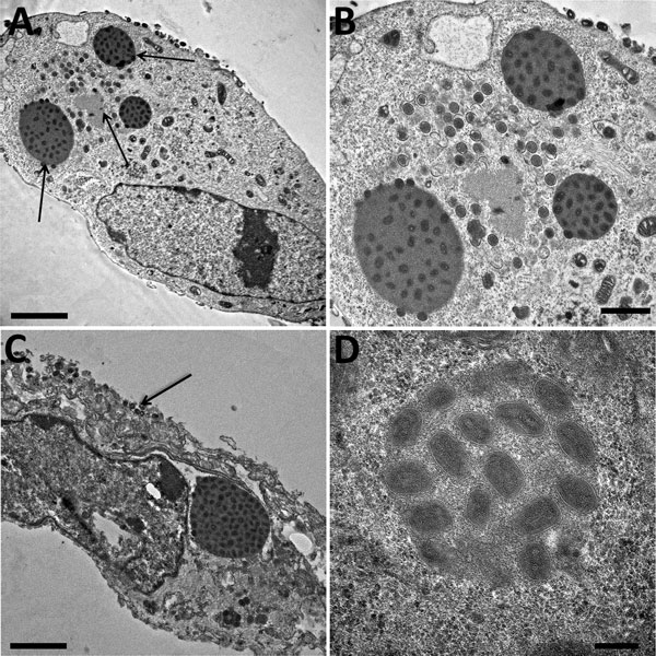
Figure 4. Electron microscopy imaging of cowpox virus France Amiens 2016, obtained from a smallpox-vaccinated patient in France in 2016. A) Ultrathin sections of a Hep2 cell at 32 hours postinfection. The cell harbors, which is undergoing its replicative cycle. Arrows indicate dense inclusion bodies as well as its viral factory containing viral crescents in the cell cytoplasm. Scale bar indicates 2 μm. B) Higher magnification of Hep2 cell in panel A; scale bar indicates 1 μm. C) Ultrathin sections of a Hep2 cell with a typical inclusion of cowpox virus detected near the nucleus. Arrow indicates extracellular-enveloped viruses or cell-associated enveloped particles. Scale bar indicates 2 μm. D) Electron-dense inclusion body containing mature viral particles. Scale bars indicate 200 nm.
Main Article
Page created: January 18, 2019
Page updated: January 18, 2019
Page reviewed: January 18, 2019
The conclusions, findings, and opinions expressed by authors contributing to this journal do not necessarily reflect the official position of the U.S. Department of Health and Human Services, the Public Health Service, the Centers for Disease Control and Prevention, or the authors' affiliated institutions. Use of trade names is for identification only and does not imply endorsement by any of the groups named above.