Volume 25, Number 4—April 2019
Synopsis
Lobomycosis in Soldiers, Colombia
Claudia M. Arenas1, Gerzain Rodriguez-Toro, Andrea Ortiz-Florez , and Ingrid Serrato
Figure 4
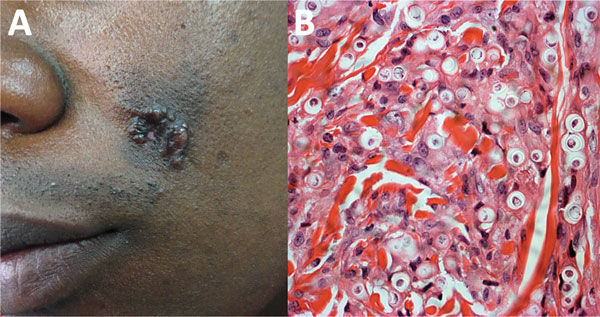
Figure 4. Lobomycosis in a 30-year-old soldier (case-patient 4), Colombia. A) Phototype V lesion on the left cheek with papules that became confluent and formed a lobulated plaque with a smooth and shiny surface. B) Periodic acid–Schiff staining of a biopsy specimen from the lesion shows multiple yeasts of uniform size, and thick walls are seen inside phagocytoses (original magnification ×40).
1Current affiliation: Hospital Universitario Centro Dermatologico Federico Lleras Acosta, Bogota, Colombia.
Page created: March 18, 2019
Page updated: March 18, 2019
Page reviewed: March 18, 2019
The conclusions, findings, and opinions expressed by authors contributing to this journal do not necessarily reflect the official position of the U.S. Department of Health and Human Services, the Public Health Service, the Centers for Disease Control and Prevention, or the authors' affiliated institutions. Use of trade names is for identification only and does not imply endorsement by any of the groups named above.