Volume 25, Number 7—July 2019
Dispatch
Diagnosis of Chagasic Encephalitis by Sequencing of 28S rRNA Gene
Ashrit Multani , Aabed Meer, Darvin S. Smith, Malika N. Kheraj, Edward D. Plowey, and Brian G. Blackburn
Figure 1
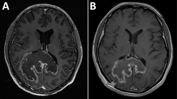
Figure 1. Images obtained during diagnosis of chagasic encephalitis in 31-year-old man in the United States. A) Contrast-enhanced T1-weighted magnetic resonance imaging of the brain showing a cerebral tumor-like chagoma in the axial plane. B) Follow-up contrast-enhanced T1-weighted magnetic resonance imaging obtained ≈8 weeks later showing improvement of the chagoma.
Page created: June 17, 2019
Page updated: June 17, 2019
Page reviewed: June 17, 2019
The conclusions, findings, and opinions expressed by authors contributing to this journal do not necessarily reflect the official position of the U.S. Department of Health and Human Services, the Public Health Service, the Centers for Disease Control and Prevention, or the authors' affiliated institutions. Use of trade names is for identification only and does not imply endorsement by any of the groups named above.