Volume 26, Number 11—November 2020
Research Letter
Nontuberculous Mycobacterial Pulmonary Disease from Mycobacterium hassiacum, Austria
Helmut J.F. Salzer , Bakari Chitechi, Doris Hillemann, Michael Mandl, Christian Paar, Monika Mitterhumer, Bernd Lamprecht, and Florian P. Maurer
Figure
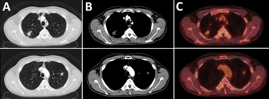
Figure. High-resolution computed tomography and 18F-fluorodeoxyglucose positron emission tomography scans of the chest showing pulmonary lesions caused by Mycobacterium hassiacum in a 62-year-old man, Austria. A and B) Computed tomography scans of the chest showing a subpleural thick-walled cavitary lesion in the posterior segment of the right upper lung lobe with associated pleural thickening and a smaller adjacent partly calcified solitary nodule. Another solid nodule of 13 mm diameter was found in the left upper lung lobe. C) Positron emission tomography scan showing a tracer uptake in both lesions with a standardized uptake values of 5 (top image) and 1.9 (bottom image).
Page created: August 18, 2020
Page updated: October 19, 2020
Page reviewed: October 19, 2020
The conclusions, findings, and opinions expressed by authors contributing to this journal do not necessarily reflect the official position of the U.S. Department of Health and Human Services, the Public Health Service, the Centers for Disease Control and Prevention, or the authors' affiliated institutions. Use of trade names is for identification only and does not imply endorsement by any of the groups named above.