Skip directly to site content Skip directly to page options Skip directly to A-Z link Skip directly to A-Z link Skip directly to A-Z link
Volume 26, Number 7—July 2020
Dispatch

Aerosol and Surface Distribution of Severe Acute Respiratory Syndrome Coronavirus 2 in Hospital Wards, Wuhan, China, 2020

Zhen-Dong Guo1, Zhong-Yi Wang1, Shou-Feng Zhang1, Xiao Li, Lin Li, Chao Li, Yan Cui, Rui-Bin Fu, Yun-Zhu Dong, Xiang-Yang Chi, Meng-Yao Zhang, Kun Liu, Cheng Cao, Bin Liu, Ke Zhang, Yu-Wei GaoComments to Author , Bing LuComments to Author , and Wei ChenComments to Author 
Author affiliations: Academy of Military Medical Sciences, Beijing, China (Z.-D. Guo, Z.-Y. Wang, S.-F. Zhang, X. Li, L. Li, Y.-Z. Dong, X.-Y. Chi, M.-Y. Zhang, C. Cao, K. Zhang, Y.-W. Gao, B. Lu, W. Chen); Institute of Medical Support Technology, Institute of Systems Engineering, Academy of Military Sciences, Tianjin, China (C. Li); Wuhan Huoshenshan Hospital, Wuhan, China (Y. Cui, R.-B. Fu, B. Liu); Central Theater General Hospital, Wuhan (K. Liu)

Main Article

Figure 2

Spatial distribution of severe acute respiratory syndrome coronavirus 2 aerosols in isolation wards of the intensive care unit (ICU) and the general ward at Huoshenshan Hospital, Wuhan, China. A) The air sampling sites in the ICU were distributed in different regions: near the air outlet (site 1), near the patients (site 2), and around the doctors’ office area (site 3). Orange circles represent sampling sites; blue arrows represent direction of the fresh air flow; and the graded orange arrow and

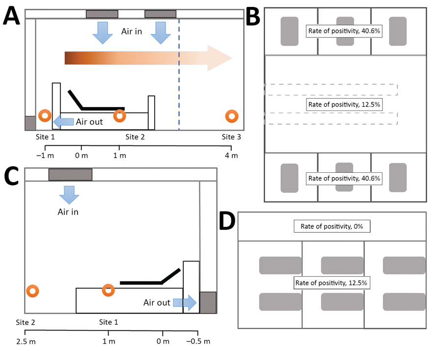
Figure 2. Spatial distribution of severe acute respiratory syndrome coronavirus 2 aerosols in isolation wards of the intensive care unit (ICU) and the general ward at Huoshenshan Hospital, Wuhan, China. A) The air sampling sites in the ICU were distributed in different regions: near the air outlet (site 1), near the patients (site 2), and around the doctors’ office area (site 3). Orange circles represent sampling sites; blue arrows represent direction of the fresh air flow; and the graded orange arrow and scale bar indicate the horizontal distance from the patient’s head. B) In terms of viral aerosol distribution, the space in the ICU was divided into 2 parts: a high-risk area with a 40.6% rate of virus positivity and a low-risk area with a 12.5% rate of virus positivity. C) The air sampling sites in the general ward were distributed in different regions around the patient (site 1), under the air inlet (site 2), and in the patient corridor. D) In terms of the viral aerosol distribution, the space in the general ward was divided into 2 parts: a high-risk area with a 12.5% rate of virus positivity and a low-risk area with a 0% rate of virus positivity.

Main Article

1These authors contributed equally to this article.

Page created: April 10, 2020
Page updated: June 18, 2020
Page reviewed: June 18, 2020
The conclusions, findings, and opinions expressed by authors contributing to this journal do not necessarily reflect the official position of the U.S. Department of Health and Human Services, the Public Health Service, the Centers for Disease Control and Prevention, or the authors' affiliated institutions. Use of trade names is for identification only and does not imply endorsement by any of the groups named above.
file_external