Volume 26, Number 9—September 2020
CME ACTIVITY - Synopsis
Q Fever Osteoarticular Infection in Children
Halima Dabaja-Younis , Michal Meir, Anat Ilivizki, Daniela Militianu, Mark Eidelman, Imad Kassis1, and Yael Shachor-Meyouhas1
Figure 1
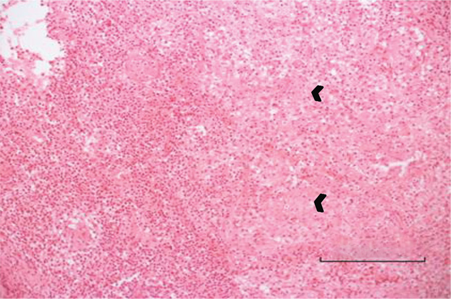
Figure 1. Bone biopsy specimen for a 3-year-old boy (case 1) with Q fever osteoarticular infection, Israel. Hematoxylin and eosin stain shows an acute inflammatory process with neutrophil and lymphocyte predominance. Small arrows indicate giant cells and epithelioid granuloma without necrosis. Bar indicates the diameter of a giant granuloma.
1These authors contributed equally to the study and article.
Page created: July 02, 2020
Page updated: August 18, 2020
Page reviewed: August 18, 2020
The conclusions, findings, and opinions expressed by authors contributing to this journal do not necessarily reflect the official position of the U.S. Department of Health and Human Services, the Public Health Service, the Centers for Disease Control and Prevention, or the authors' affiliated institutions. Use of trade names is for identification only and does not imply endorsement by any of the groups named above.