Volume 26, Number 9—September 2020
Dispatch
Hepatitis E Virus Genotype 7 RNA and Antibody Kinetics in Naturally Infected Dromedary Calves, United Arab Emirates
Victor M. Corman , Peter Nagy, Stefanie Ostermann, Jacqueline Arloth, Anne Liljander, Rajib Barua, Aungshuman Das Gupta, Fatima Hakimuddin, Judit Juhasz, Ulrich Wernery, and Christian Drosten
Figure 1
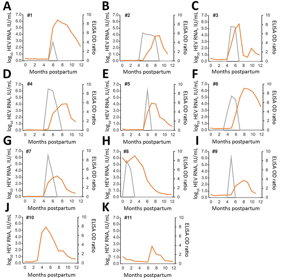
Figure 1. HEV RNA and HEV antibodies in 11 dromedary calves, United Arab Emirates. Gray lines indicate HEV RNA concentration in serum, and orange lines indicate antibody levels. HEV, hepatitis E virus; OD, optical density.
Page created: May 04, 2020
Page updated: August 19, 2020
Page reviewed: August 19, 2020
The conclusions, findings, and opinions expressed by authors contributing to this journal do not necessarily reflect the official position of the U.S. Department of Health and Human Services, the Public Health Service, the Centers for Disease Control and Prevention, or the authors' affiliated institutions. Use of trade names is for identification only and does not imply endorsement by any of the groups named above.