Skip directly to site content Skip directly to page options Skip directly to A-Z link Skip directly to A-Z link Skip directly to A-Z link
Volume 27, Number 11—November 2021
Research

Encephalitis and Death in Wild Mammals at a Rehabilitation Center after Infection with Highly Pathogenic Avian Influenza A(H5N8) Virus, United Kingdom

Tobias Floyd, Ashley C. BanyardComments to Author , Fabian Z.X. Lean, Alexander M.P. Byrne, Edward Fullick, Elliot Whittard, Benjamin C. Mollett, Steve Bexton, Vanessa Swinson, Michele Macrelli, Nicola S. Lewis, Scott M. Reid, Alejandro Núñez, J. Paul Duff, Rowena Hansen, and Ian H. Brown
Author affiliations: Animal and Plant Health Agency, Weybridge, UK (T. Floyd, A.C. Banyard, F.Z.X Lean, A.M.P. Byrne, E. Whittard, B.C. Mollett, S.M. Reid, A. Núñez, R. Hansen, I.H. Brown); Animal and Plant Health Agency, Thirsk, UK (E. Fullick, V. Swinson); Royal Society for Prevention of Cruelty to Animals, East Winch, UK (S. Bexton); Animal and Plant Health Agency, Bury St Edmunds, UK (M. Macrelli); Royal Veterinary College Department of Pathobiology and Population Sciences, North Mymms, UK (N.S. Lewis); Animal and Plant Health Agency Diseases of Wildlife Scheme, Penrith, UK (J.P. Duff)

Main Article

Figure 2

Histopathologic and immunohistochemical examination of the gray seal (Halichoerus grypus), common seal (Phoca vitulina), and red fox (Vulpes vulpes) infected with highly pathogenic avian influenza A subtype H5N8, United Kingdom. Serial tissue sections were stained with hematoxylin and eosin. Immunohistochemical examination was undertaken using anti–influenza A nucleoprotein primary antibody (Statens Serum Institute, https://en.ssi.dk). Insets show histopathologic study results. A) Nonsuppurative polioencephalitis and presence of virus antigens in neurons in the cerebrum, common seal (Phoca vitulina). Original magnification ×10, inset ×40. B) Nonsuppurative polioencephalitis with neuronophagia and association of virus antigens, red fox. Original magnification ×10, inset ×40. C) Ependymal necrosis and the association of virus antigens, fox. Original magnification and inset ×40; area of interest also shown. D) Diffuse alveolar damage and presence of virus in type I alveolar pneumocytes, red fox. Original magnification and inset ×40. E) Cardiomyonecrosis associated with virus antigens in cardiomyocytes, red fox. Original magnification and inset ×40. F) Virus antigens in granular and molecular layer of the cerebellum, red fox. Original magnification ×20, inset ×40. Serial tissue sections were stained with hematoxylin and eosin. Immunohistochemical examination was undertaken using anti–influenza A nucleoprotein primary antibody (Statens Serum Institute, https://en.ssi.dk). Insets show histopathologic study results.

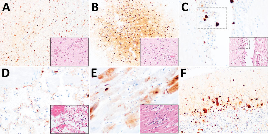
Figure 2. Histopathologic and immunohistochemical examination of the gray seal (Halichoerus grypus), common seal (Phoca vitulina), and red fox (Vulpes vulpes) infected with highly pathogenic avian influenza A subtype H5N8, United Kingdom. Serial tissue sections were stained with hematoxylin and eosin. Immunohistochemical examination was undertaken using anti–influenza A nucleoprotein primary antibody (Statens Serum Institute, https://en.ssi.dk). Insets show histopathologic study results. A) Nonsuppurative polioencephalitis and presence of virus antigens in neurons in the cerebrum, common seal (Phoca vitulina). Original magnification ×10, inset ×40. B) Nonsuppurative polioencephalitis with neuronophagia and association of virus antigens, red fox. Original magnification ×10, inset ×40. C) Ependymal necrosis and the association of virus antigens, fox. Original magnification and inset ×40; area of interest also shown. D) Diffuse alveolar damage and presence of virus in type I alveolar pneumocytes, red fox. Original magnification and inset ×40. E) Cardiomyonecrosis associated with virus antigens in cardiomyocytes, red fox. Original magnification and inset ×40. F) Virus antigens in granular and molecular layer of the cerebellum, red fox. Original magnification ×20, inset ×40. Serial tissue sections were stained with hematoxylin and eosin. Immunohistochemical examination was undertaken using anti–influenza A nucleoprotein primary antibody (Statens Serum Institute, https://en.ssi.dk). Insets show histopathologic study results.

Main Article

Page created: September 13, 2021
Page updated: October 19, 2021
Page reviewed: October 19, 2021
The conclusions, findings, and opinions expressed by authors contributing to this journal do not necessarily reflect the official position of the U.S. Department of Health and Human Services, the Public Health Service, the Centers for Disease Control and Prevention, or the authors' affiliated institutions. Use of trade names is for identification only and does not imply endorsement by any of the groups named above.
file_external