Surface‒Aerosol Stability and Pathogenicity of Diverse Middle East Respiratory Syndrome Coronavirus Strains, 2012‒2018
Neeltje van Doremalen
1, Michael Letko
1
, Robert J. Fischer, Trenton Bushmaker, Jonathan Schulz, Claude K. Yinda, Stephanie N. Seifert, Nam Joong Kim, Maged G. Hemida, Ghazi Kayali, Wan Beom Park, Ranawaka A.P.M. Perera, Azaibi Tamin, Natalie J. Thornburg, Suxiang Tong, Krista Queen, Maria D. van Kerkhove, Young Ki Choi, Myoung-don Oh, Abdullah M. Assiri, Malik Peiris, Susan I. Gerber, and Vincent J. Munster
Author affiliations: National Institutes of Health, Hamilton, Montana, USA (N. van Doremalen, M. Letko, R.J. Fischer, T. Bushmaker, J. Schulz, C.K. Yinda, S.N. Seifert, V.J. Munster); Washington State University, Pullman, Washington, USA (M. Leitko, S.N. Seifert); Seoul National University College of Medicine, Seoul, South Korea (N.J. Kim, W.B. Park, M.-d. Oh); King Faisal University, Al-Hasa, Saudi Arabia (M.G. Hemida); Kafrelsheikh University, Kafrelsheikh, Egypt (M.G. Hemida); University of Texas Health Sciences Center, Houston, Texas, USA (G. Kayali); University of Hong Kong, Hong Kong, China (R.A.P.M. Perera, M. Peiris); Centers for Disease Control and Prevention, Atlanta, Georgia, USA (A. Tamin, N.J. Thornburg, S. Tong, K. Queen, S.I. Gerber); World Health Organization, Geneva, Switzerland (M.D. van Kerkhove); Chungbuk National University, Cheongju City, South Korea (Y.K. Choi); Ministry of Health, Riyadh, Saudi Arabia (A.M. Assiri)
Main Article
Figure 3
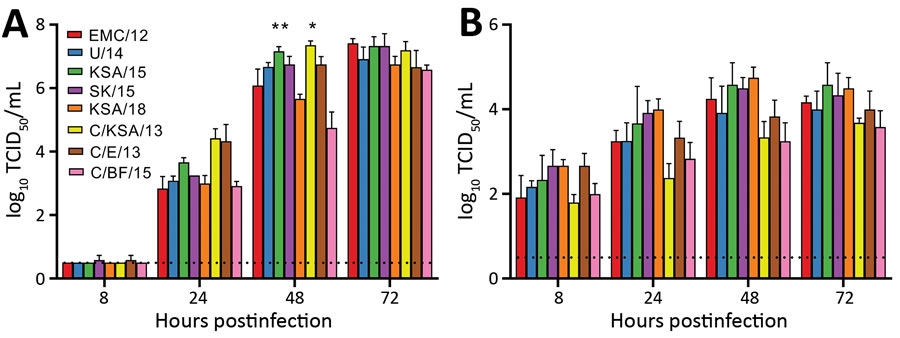
Figure 3. Middle East respiratory syndrome coronavirus replication in Vero E6 cells (A) and human airway epithelium (B). Replication is shown as geometric means; error bars indicate SDs. Vero E6 cells were infected with a multiplicity of infection of 0.01, and human airway epithelium were infected with a multiplicity of infection of 0.1. Samples of supernatants were obtained at 8, 24, 48 and 72 hours postinoculation and titrated. Statistically significant differences compared with those for the prototypical strain, EMC/12, were calculated by using ordinary 1-way analysis of variance, followed by a Bonferroni multiple comparisons test. Dotted lines indicate limits of detection. Strain sources are listed in the legend for Figure 1. TCID50, median tissue culture infectious dose. *p<0.05; **p<0.01.
Main Article
Page created: August 26, 2021
Page updated: November 19, 2021
Page reviewed: November 19, 2021
The conclusions, findings, and opinions expressed by authors contributing to this journal do not necessarily reflect the official position of the U.S. Department of Health and Human Services, the Public Health Service, the Centers for Disease Control and Prevention, or the authors' affiliated institutions. Use of trade names is for identification only and does not imply endorsement by any of the groups named above.