Skip directly to site content Skip directly to page options Skip directly to A-Z link Skip directly to A-Z link Skip directly to A-Z link
Volume 27, Number 2—February 2021
Research

Prolonged Maternal Zika Viremia as a Marker of Adverse Perinatal Outcomes

Léo Pomar, Véronique Lambert, Séverine Matheus, Céline Pomar, Najeh Hcini, Gabriel Carles, Dominique Rousset, Manon Vouga, Alice Panchaud1, and David Baud1Comments to Author 
Author affiliations: Lausanne University Hospital, Lausanne, Switzerland (L. Pomar, C. Pomar, M. Vouga, A. Panchaud, D. Baud); Centre Hospitalier de l’Ouest Guyanais Franck Joly, Saint-Laurent-du-Maroni, French Guiana (V. Lambert, C. Pomar, N. Hcini, G. Carles); Institut Pasteur, Paris, France (S. Matheus); Institut Pasteur of French Guiana, Cayenne, French Guiana (S. Matheus, D. Rousset); University of Bern, Bern, Switzerland (A. Panchaud)

Main Article

Figure 3

Prenatal ultrasound, computed tomography, and postmortem aspect of a fetus with congenital Zika syndrome related to maternal prolonged viremia in patient (case no. 122) in a cohort study of pregnant women admitted to Centre Hospitalier de l’Ouest Guyanais, French Guiana, January 1–July 15, 2016. The mother had symptomatic acute Zika virus infection at 8 weeks’ gestation (and had ongoing viremia until birth of her stillborn child with signs of congenital Zika syndrome. Severe microcephaly, ventriculomegaly, and calcifications were detected by ultrasound at 13 weeks’ gestation. Overall, this fetus had arthrogryposis detected on 3-D ultrasound (A) and postmortem (B); severe bilateral microphthalmia (blue arrows) detected on 3-D ultrasound (C) and fetal computed tomography (D); microcephaly with atrophic cortex detected on ultrasound (E) showing a head circumference of 160 mm at 25 weeks’ gestation (−5 SDs); ventriculomegaly detected on ultrasound (E); brain calcifications (blue arrows) detected on ultrasound (F) and computed tomography (G); pontocerebellar hypoplasia (yellow arrows) detected on ultrasound (F); and corpus callosum dysgenesis (yellow arrows) detected on ultrasound (H).

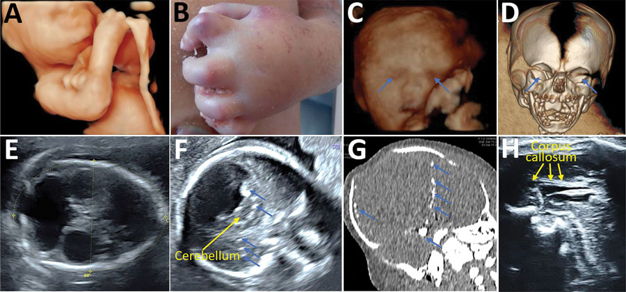
Figure 3. Prenatal ultrasound, computed tomography, and postmortem aspect of a fetus with congenital Zika syndrome related to maternal prolonged viremia in patient (case no. 122) in a cohort study of pregnant women admitted to Centre Hospitalier de l’Ouest Guyanais, French Guiana, January 1–July 15, 2016. The mother had symptomatic acute Zika virus infection at 8 weeks’ gestation (and had ongoing viremia until birth of her stillborn child with signs of congenital Zika syndrome. Severe microcephaly, ventriculomegaly, and calcifications were detected by ultrasound at 13 weeks’ gestation. Overall, this fetus had arthrogryposis detected on 3-D ultrasound (A) and postmortem (B); severe bilateral microphthalmia (blue arrows) detected on 3-D ultrasound (C) and fetal computed tomography (D); microcephaly with atrophic cortex detected on ultrasound (E) showing a head circumference of 160 mm at 25 weeks’ gestation (−5 SDs); ventriculomegaly detected on ultrasound (E); brain calcifications (blue arrows) detected on ultrasound (F) and computed tomography (G); pontocerebellar hypoplasia (yellow arrows) detected on ultrasound (F); and corpus callosum dysgenesis (yellow arrows) detected on ultrasound (H).

Main Article

1These senior authors contributed equally to this article.

Page created: December 10, 2020
Page updated: January 23, 2021
Page reviewed: January 23, 2021
The conclusions, findings, and opinions expressed by authors contributing to this journal do not necessarily reflect the official position of the U.S. Department of Health and Human Services, the Public Health Service, the Centers for Disease Control and Prevention, or the authors' affiliated institutions. Use of trade names is for identification only and does not imply endorsement by any of the groups named above.
file_external