Skip directly to site content Skip directly to page options Skip directly to A-Z link Skip directly to A-Z link Skip directly to A-Z link
Volume 27, Number 2—February 2021
Research Letter

Protective Immunity and Persistent Lung Sequelae in Domestic Cats after SARS-CoV-2 Infection

Shiho Chiba, Peter J. Halfmann, Masato Hatta, Tadashi Maemura, Shufang Fan, Tammy Armbrust, Olivia M. Swartley, LaTasha K. CrawfordComments to Author , and Yoshihiro KawaokaComments to Author 
Author affiliations: University of Wisconsin–Madison School of Veterinary Medicine, Madison, Wisconsin, USA (S. Chiba, P.J. Halfmann, M. Hatta, T. Maemura, S. Fan, T. Armbrust, O.M. Swartley, L.K. Crawford, Y. Kawaoka,); University of Tokyo Institute of Medical Science, Tokyo, Japan (Y. Kawaoka)

Main Article

Figure 2

Timeline of severe acute respiratory syndrome coronavirus 2 infection and reinfection of cats and distribution of interstitial thickening. A) Timeline of infection and reinfection. As reported previously (1), a group of cats was inoculated with severe acute respiratory syndrome coronavirus 2 on day 0 (infected cats 1–3, upper half). A virus-naive cat was cohoused with each of the infected cats from day 1 (contact cats 1–3, lower half). The days on which infectious virus was detected in the nasal swabs are shown as red bars for each animal. In this study, we infected the cats with the same severe acute respiratory syndrome coronavirus 2 isolate at ≈4 weeks after initial infection or exposure to infected cats. After reinfection of the group shown in the upper half of the figure, no infectious virus was detected in the nasal swabs. The cats were confirmed to be seronegative before the initial infection or cohousing with infected cats, and seropositive before reinfection, on the basis of neutralization assay results. B) The distribution of interstitial thickening (interstitial pneumonia severity score) was decreased on day 21 after reinfection compared with day 28 (p = 0.041 by unpaired t-test).

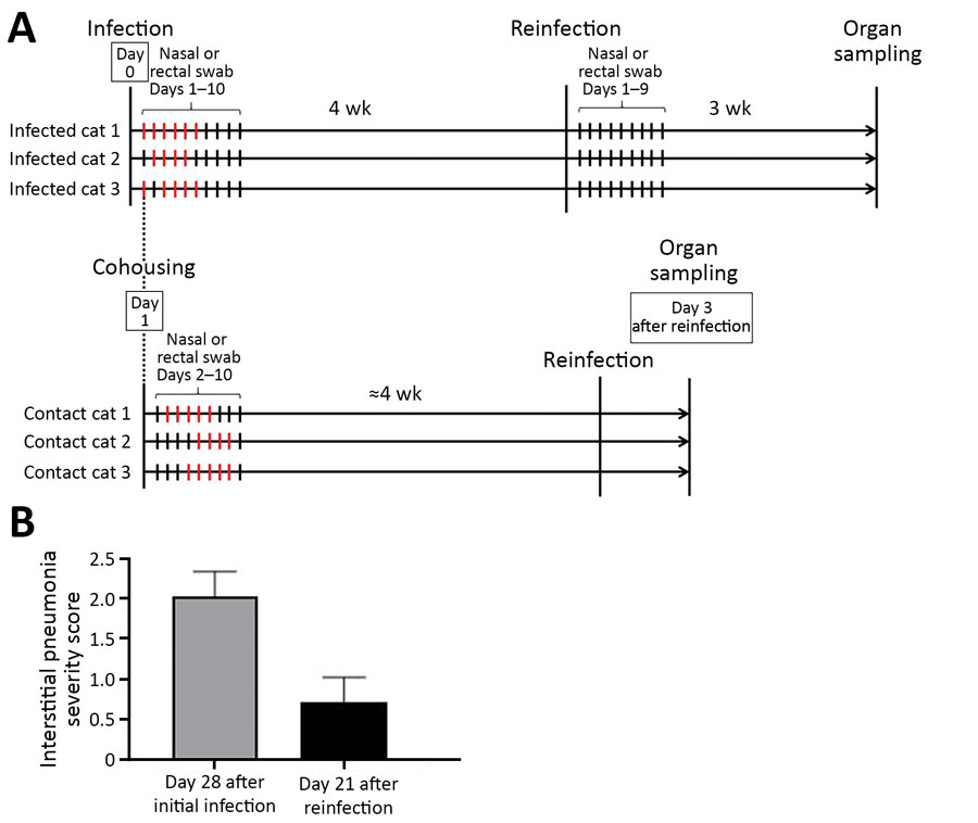
Figure 2. Timeline of severe acute respiratory syndrome coronavirus 2 infection and reinfection of cats and distribution of interstitial thickening. A) Timeline of infection and reinfection. As reported previously (1), a group of cats was inoculated with severe acute respiratory syndrome coronavirus 2 on day 0 (infected cats 1–3, upper half). A virus-naive cat was cohoused with each of the infected cats from day 1 (contact cats 1–3, lower half). The days on which infectious virus was detected in the nasal swabs are shown as red bars for each animal. In this study, we infected the cats with the same severe acute respiratory syndrome coronavirus 2 isolate at ≈4 weeks after initial infection or exposure to infected cats. After reinfection of the group shown in the upper half of the figure, no infectious virus was detected in the nasal swabs. The cats were confirmed to be seronegative before the initial infection or cohousing with infected cats, and seropositive before reinfection, on the basis of neutralization assay results. B) The distribution of interstitial thickening (interstitial pneumonia severity score) was decreased on day 21 after reinfection compared with day 28 (p = 0.041 by unpaired t-test).

Main Article

References
  1. Halfmann  PJ, Hatta  M, Chiba  S, Maemura  T, Fan  S, Takeda  M, et al. Transmission of SARS-CoV-2 in domestic cats. N Engl J Med. 2020;383:5924. DOIPubMedGoogle Scholar
  2. Shi  J, Wen  Z, Zhong  G, Yang  H, Wang  C, Huang  B, et al. Susceptibility of ferrets, cats, dogs, and other domesticated animals to SARS-coronavirus 2. Science. 2020;368:101620. DOIPubMedGoogle Scholar
  3. Cheung  OY, Chan  JW, Ng  CK, Koo  CK. The spectrum of pathological changes in severe acute respiratory syndrome (SARS). Histopathology. 2004;45:11924. DOIPubMedGoogle Scholar
  4. Das  KM, Lee  EY, Singh  R, Enani  MA, Al Dossari  K, Van Gorkom  K, et al. Follow-up chest radiographic findings in patients with MERS-CoV after recovery. Indian J Radiol Imaging. 2017;27:3429. DOIPubMedGoogle Scholar
  5. Ackermann  M, Verleden  SE, Kuehnel  M, Haverich  A, Welte  T, Laenger  F, et al. Pulmonary vascular endothelialitis, thrombosis, and angiogenesis in Covid-19. N Engl J Med. 2020;383:1208. DOIPubMedGoogle Scholar

Main Article

Page created: November 09, 2020
Page updated: January 24, 2021
Page reviewed: January 24, 2021
The conclusions, findings, and opinions expressed by authors contributing to this journal do not necessarily reflect the official position of the U.S. Department of Health and Human Services, the Public Health Service, the Centers for Disease Control and Prevention, or the authors' affiliated institutions. Use of trade names is for identification only and does not imply endorsement by any of the groups named above.
file_external