Skip directly to site content Skip directly to page options Skip directly to A-Z link Skip directly to A-Z link Skip directly to A-Z link
Volume 27, Number 4—April 2021
Research

Histopathological Characterization of Cases of Spontaneous Fatal Feline Severe Fever with Thrombocytopenia Syndrome, Japan

Yusuke SakaiComments to Author , Yuko Kuwabara, Keita Ishijima1, Saya Kagimoto, Serina Mura, Kango Tatemoto1, Ryusei Kuwata2, Kenzo Yonemitsu1, Shohei Minami, Yudai Kuroda1, Kenji Baba, Masaru Okuda, Hiroshi Shimoda, Masashi Sakurai, Masahiro Morimoto, and Ken Maeda1
Author affiliation: Yamaguchi University, Yamaguchi, Japan

Main Article

Figure 3

Histopathological lesions in the intestinal tracts from fatal cases of severe fever with thrombocytopenia syndrome (SFTS) in cats, Japan. A, B) Hematoxylin & eosin (HE)–stained (B) and immunohistochemistry-stained (A) ileum sections demonstrating enlargement of Peyer’s patch and accumulation of SFTSV-positive blastic lymphocytes. Scale bars indicates 100 μm. C) HE-stained colon sections demonstrating infiltration of lymphocytes into the lamina propria. Scale bar indicates 100 μm. D) High power magnification of panel C demonstrating the infiltrating lymphocytes were blastic lymphocytes. Scale bar indicates 10 μm. E, F) HE stained (E) and immunohistochemistry-stained (F) ulcerative lesions in the cecum. Scale bars indicate 200 μm.

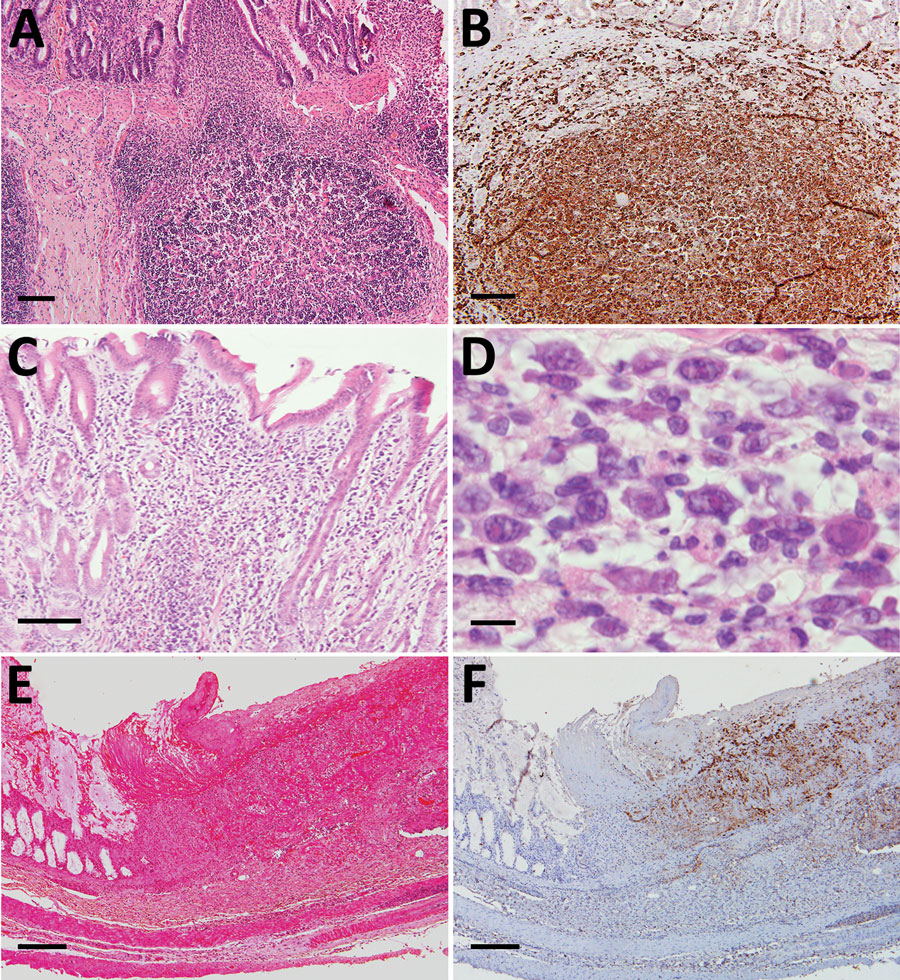
Figure 3. Histopathological lesions in the intestinal tracts from fatal cases of severe fever with thrombocytopenia syndrome (SFTS) in cats, Japan. A, B) Hematoxylin & eosin (HE)–stained (B) and immunohistochemistry-stained (A) ileum sections demonstrating enlargement of Peyer’s patch and accumulation of SFTSV-positive blastic lymphocytes. Scale bars indicates 100 μm. C) HE-stained colon sections demonstrating infiltration of lymphocytes into the lamina propria. Scale bar indicates 100 μm. D) High power magnification of panel C demonstrating the infiltrating lymphocytes were blastic lymphocytes. Scale bar indicates 10 μm. E, F) HE stained (E) and immunohistochemistry-stained (F) ulcerative lesions in the cecum. Scale bars indicate 200 μm.

Main Article

1Current affiliation: National Institute of Infectious Diseases, Tokyo, Japan.

2Current affiliation: Okayama University of Science, Ehime, Japan.

Page created: February 17, 2021
Page updated: March 18, 2021
Page reviewed: March 18, 2021
The conclusions, findings, and opinions expressed by authors contributing to this journal do not necessarily reflect the official position of the U.S. Department of Health and Human Services, the Public Health Service, the Centers for Disease Control and Prevention, or the authors' affiliated institutions. Use of trade names is for identification only and does not imply endorsement by any of the groups named above.
file_external