Skip directly to site content Skip directly to page options Skip directly to A-Z link Skip directly to A-Z link Skip directly to A-Z link
Volume 27, Number 4—April 2021
Synopsis

Difficulties in Differentiating Coronaviruses from Subcellular Structures in Human Tissues by Electron Microscopy

Hannah A. BullockComments to Author , Cynthia S. Goldsmith, Sherif R. Zaki, Roosecelis B. Martines, and Sara E. Miller
Author affiliations: Synergy America, Inc., Atlanta, Georgia, USA (H.A. Bullock); Centers for Disease Control and Prevention, Atlanta (C.S. Goldsmith, S.R. Zaki, R.B. Martines); Duke Medical Center, Durham, North Carolina, USA (S.E. Miller)

Main Article

Figure 1

Overview of the ultrastructural features of coronavirus morphology as seen by negative stain and thin section. A) Extracellular viral particles ≈100 nm in diameter with prominent peplomers (spikes). Prepared from a cell culture sample by negative stain using heavy metal salt solutions to coat the outside of the virus. Scale bar indicates 100 nm. B) Extracellular viral particles ≈100 nm in diameter with clearly visible spikes. Cross sections through the helical nucleocapsid are visible on the interior of the particle as electron-dense black dots, 6–12 nm in diameter. Prepared by thin section from a formalin-fixed autopsy specimen. Scale bar indicates 100 nm. C) Intracellular viral particles ≈80 nm in diameter held within a membrane-bound vacuole. Cross sections through the helical nucleocapsid are visible inside the particles. Prepared by thin section from a formalin-fixed autopsy specimen. Scale bar indicates 200 nm. D) Intracellular viral particles (arrowhead) within a membrane-bound vacuole and nearby clathrin-coated vesicles (CCV) in the cytoplasm (arrows). CCV spikes directly contact the cell cytosol; viral spikes, barely visible as a faint fuzz, contact the vacuole contents. Cross sections through the helical nucleocapsid are visible inside the viral particles but not within the CCVs. Prepared by thin section from a glutaraldehyde-fixed cell culture sample. Scale bar indicates 500 nm.

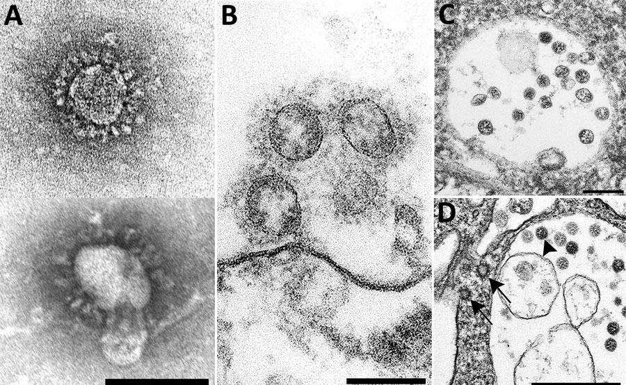
Figure 1. Overview of the ultrastructural features of coronavirus morphology as seen by negative stain and thin section. A) Extracellular viral particles ≈100 nm in diameter with prominent peplomers (spikes). Prepared from a cell culture sample by negative stain using heavy metal salt solutions to coat the outside of the virus. Scale bar indicates 100 nm. B) Extracellular viral particles ≈100 nm in diameter with clearly visible spikes. Cross sections through the helical nucleocapsid are visible on the interior of the particle as electron-dense black dots, 6–12 nm in diameter. Prepared by thin section from a formalin-fixed autopsy specimen. Scale bar indicates 100 nm. C) Intracellular viral particles ≈80 nm in diameter held within a membrane-bound vacuole. Cross sections through the helical nucleocapsid are visible inside the particles. Prepared by thin section from a formalin-fixed autopsy specimen. Scale bar indicates 200 nm. D) Intracellular viral particles (arrowhead) within a membrane-bound vacuole and nearby clathrin-coated vesicles (CCV) in the cytoplasm (arrows). CCV spikes directly contact the cell cytosol; viral spikes, barely visible as a faint fuzz, contact the vacuole contents. Cross sections through the helical nucleocapsid are visible inside the viral particles but not within the CCVs. Prepared by thin section from a glutaraldehyde-fixed cell culture sample. Scale bar indicates 500 nm.

Main Article

Page created: February 18, 2021
Page updated: March 18, 2021
Page reviewed: March 18, 2021
The conclusions, findings, and opinions expressed by authors contributing to this journal do not necessarily reflect the official position of the U.S. Department of Health and Human Services, the Public Health Service, the Centers for Disease Control and Prevention, or the authors' affiliated institutions. Use of trade names is for identification only and does not imply endorsement by any of the groups named above.
file_external