Volume 27, Number 5—May 2021
Research Letter
Eosinophilic Meningitis and Intraocular Infection Caused by Dirofilaria sp. Genotype Hongkong
Aruna S. Jyotsna, Kollencheri Puthenveettil Vinayan , Lalitha Biswas, Sujithra Haridas, Arun G. Roy, Parasmal Suresh, and Anil Kumar
Figure
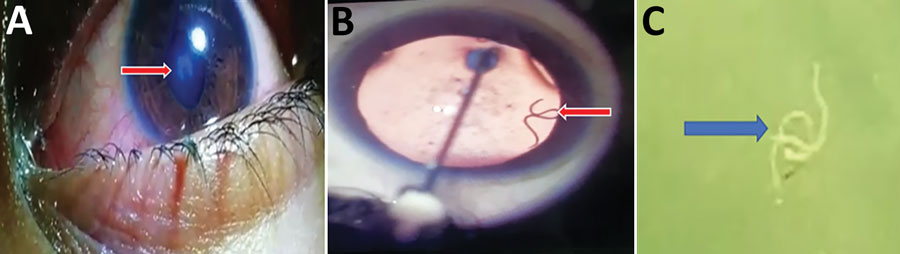
Figure. Eosinophilic meningitis and intraocular infection caused by Dirofilaria sp. genotype Hongkong in a patient in Kochi, Inida. A) Organism (arrow) in the left eye of patient during routine clinical examination. The organism caused an abnormal shape of the pupil. B) Live worm (arrow) in anterior chamber of the left eye. This image was obtained while lignocaine was being injected. C) Gross specimen of the worm (arrow) after extraction. Worm is in saline in a Petri dish.
Page created: February 24, 2021
Page updated: April 21, 2021
Page reviewed: April 21, 2021
The conclusions, findings, and opinions expressed by authors contributing to this journal do not necessarily reflect the official position of the U.S. Department of Health and Human Services, the Public Health Service, the Centers for Disease Control and Prevention, or the authors' affiliated institutions. Use of trade names is for identification only and does not imply endorsement by any of the groups named above.