Susceptibility of Well-Differentiated Airway Epithelial Cell Cultures from Domestic and Wild Animals to Severe Acute Respiratory Syndrome Coronavirus 2
Mitra Gultom, Matthias Licheri, Laura Laloli, Manon Wider, Marina Strässle, Philip V’kovski
1, Silvio Steiner, Annika Kratzel, Tran Thi Nhu Thao, Lukas Probst, Hanspeter Stalder, Jasmine Portmann, Melle Holwerda, Nadine Ebert, Nadine Stokar-Regenscheit, Corinne Gurtner, Patrik Zanolari, Horst Posthaus, Simone Schuller, Amanda Vicente-Santos, Andres Moreira-Soto, Eugenia Corrales-Aguilar, Nicolas Ruggli, Gergely Tekes, Veronika von Messling, Bevan Sawatsky, Volker Thiel, and Ronald Dijkman

Author affiliations: Institute for Infectious Diseases, University of Bern, Bern, Switzerland (M. Gultom, M. Licheri, L. Laloli, M. Wider, M. Strässle, L. Probst, M. Holwerda, R. Dijkman); University of Bern Department of Infectious Diseases and Pathobiology, Bern (M. Gultom, L. Laloli, M. Strässle, P. V’kovski, S. Steiner, A. Kratzel, T.T.N. Thao, H. Stalder, J. Portmann, M. Holwerda, N. Ebert, N. Stokar-Regenscheit, C. Gurtner, H. Posthaus, V. Thiel, R. Dijkman); Institute of Virology and Immunology, Bern and Mittelhäusern, Switzerland (M. Gultom, L. Laloli, M. Strässle, P. V’kovski, S. Steiner, A. Kratzel, T.T.N. Thao, H. Stalder, J. Portmann, M. Holwerda, N. Ebert, V. Thiel, R. Dijkman); University of Bern Graduate School for Biomedical Science, Bern (M. Gultom, L. Laloli, S. Steiner, A. Kratzel, T.T.N. Thao, L. Probst, M. Holwerda); Institute of Veterinary Bacteriology, University of Bern, Bern (M. Strässle); Institute of Animal Pathology, University of Bern, Bern (N. Stokar-Regenscheit, C. Gurtner, H. Posthaus); Clinic for Ruminants, Vetsuisse Faculty, University of Bern, Bern (P. Zanolari); University of Bern Department of Clinical Veterinary Medicine, Bern (S. Schuller); Virology-Research Center for tropical diseases (CIET), University of Costa Rica, Montes de Oca, Costa Rica (A. Vicente-Santos, A. Moreira-Soto, E. Corrales-Aguilar); Institute of Virology, Charité-Universitätsmedizin Berlin, Corporate Member of Freie Universität Berlin, Humboldt-Universität zu Berlin, and Berlin Institute of Health, Berlin, Germany (A. Moreira-Soto); Institute of Virology, Justus Liebig University Giessen, Giessen, Germany (G. Tekes); Federal Institute for Vaccines and Biomedicines, Langen,; Germany (V. von Messling, B. Sawatsky)
Main Article
Figure 2
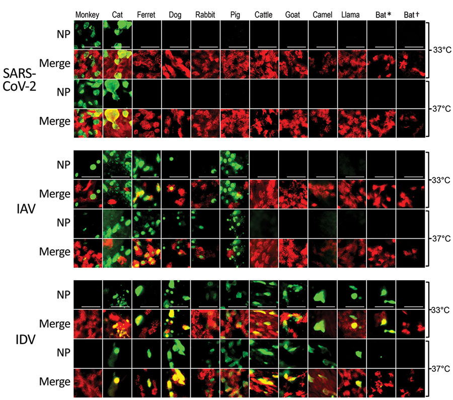
Figure 2. Tropisms of SARS-CoV-2, IAV, and IDV in infected airway epithelial cell cultures from diverse mammal species. We inoculated well-differentiated animal airway epithelial cell cultures with either 30.000 PFU of SARS-CoV-2 (SARS-CoV-2/München-1.1/2020/929), 10.000 50% tissue culture infective dose of IAV/Hamburg/4/2009, or IDV (D/bovine/Oklahoma/660/2013). We incubated virus-infected airway epithelial cell cultures at 33°C or 37°C and formalin-fixed them at 96 hours postinfection (for SARS-CoV-2) or 48 hours postinfection (for influenza viruses). After fixation, we stained virus-infected cultures using antibodies against either SARS-CoV-2, IAV, or IDV NP (green), and β-tubulin (cilia, red). We acquired images using an EVOS FL Auto 2 Imaging System equipped with a 40x air objective. Scale bar indicates 50 µm. *Sturnira lilium bat; †Carollia perspicillata bat. IAV, influenza A virus; IDV, influenza D virus; NP, nucleocapsid protein; SARS-CoV-2, severe acute respiratory syndrome coronavirus 2.
Main Article
Page created: June 16, 2021
Page updated: June 16, 2021
Page reviewed: June 16, 2021
The conclusions, findings, and opinions expressed by authors contributing to this journal do not necessarily reflect the official position of the U.S. Department of Health and Human Services, the Public Health Service, the Centers for Disease Control and Prevention, or the authors' affiliated institutions. Use of trade names is for identification only and does not imply endorsement by any of the groups named above.