Multicenter Epidemiologic Study of Coronavirus Disease–Associated Mucormycosis, India
Atul Patel
1, Ritesh Agarwal
12, Shivaprakash M. Rudramurthy, Manoj Shevkani, Immaculata Xess, Ratna Sharma, Jayanthi Savio, Nandini Sethuraman, Surabhi Madan, Prakash Shastri, Deepak Thangaraju, Rungmei Marak, Karuna Tadepalli, Pratik Savaj, Ayesha Sunavala, Neha Gupta, Tanu Singhal, Valliappan Muthu, Arunaloke Chakrabarti
2
, and
MucoCovi Network3
Author affiliations: Sterling Hospital, Ahmedabad, India (A Patel); Postgraduate Institute of Medical Education & Research, Chandigarh, India (R. Agarwal, S.M. Rudramurthy, V. Muthu, A. Chakrabarti); Avaron Hospital, Ahmedabad (M. Shevkani); All India Institute of Medical Science, New Delhi, India (I. Xess); Apollo Hospital, Hyderabad, India (R. Sharma); St. John’s Medical College, Bengaluru, India (J. Savio); Apollo Hospital, Chennai, India (N. Sethuraman); Care Institute of Medical Sciences, Ahmedabad (S. Madan); Sir Ganga Ram Hospital, New Delhi (P. Shastri); Kovai Medical Centre and Hospital, Coimbatore, India (D. Thangaraju); Sanjay Gandhi Postgraduate Institute of Medical Sciences, Lucknow, India (R. Marak); All India Institute of Medical Sciences, Bhopal, India (K. Tadepalli); Venus Hospital, Surat, India (P. Savaj); Hinduja Hospital, Mumbai, Maharashtra, India (A. Sunavala); Medanta The Medicity, Gurgaon, India (N. Gupta); Kokilaben Hospital, Mumbai, Maharashtra, India (T. Singhal)
Main Article
Figure 3
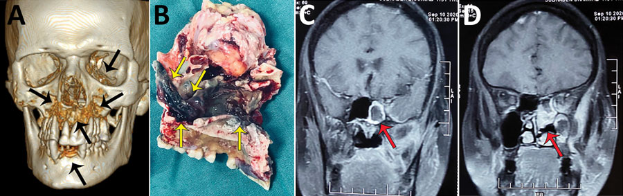
Figure 3. Radiographic images and surgical specimens demonstrating rhino-orbital-cerebral coronavirus disease–associated mucormycosis in patients from India, 2020. A) Three-dimensional reconstruction of computed tomography scan of 54-year-old male patient. Black arrows indicate patchy osteonecrosis involving the upper jaw, right orbital wall, and paranasal sinuses. B) Surgical specimen from the maxilla of 54-year-old male patient showing black necrotic paranasal sinus with palatal involvement indicated by yellow arrows. C, D) Magnetic resonance imaging (MRI) of coronal section of paranasal sinus and brain of 51-year-old female patient. Red arrow in panel C indicates enhancing cavernous sinus lesion; D) red arrow in panel D indicates right ethmoid and maxillary sinusitis. Scale bar indicates 7 cm.
Main Article
Page created: June 02, 2021
Page updated: August 17, 2021
Page reviewed: August 17, 2021
The conclusions, findings, and opinions expressed by authors contributing to this journal do not necessarily reflect the official position of the U.S. Department of Health and Human Services, the Public Health Service, the Centers for Disease Control and Prevention, or the authors' affiliated institutions. Use of trade names is for identification only and does not imply endorsement by any of the groups named above.