Skip directly to site content Skip directly to page options Skip directly to A-Z link Skip directly to A-Z link Skip directly to A-Z link
Volume 28, Number 1—January 2022
Dispatch

Streptococcus gallolyticus and Bacterial Endocarditis in Swine, United States, 2015–2020

Panchan SitthicharoenchaiComments to Author , Eric R. Burrough, Bailey L. Arruda, Orhan Sahin, Jessica G. dos Santos, Drew R. Magstadt, Pablo E. Piñeyro, Kent J. Schwartz, and Michael C. Rahe
Author affiliations: Iowa State University, Ames, Iowa, USA (P. Sitthicharoenchai, E.R. Burrough, O. Sahin, J.G. dos Santos, D.R. Magstadt, P.E. Piñeyro, K.J. Schwartz, M.C. Rahe); US Department of Agriculture, Ames (B.L. Arruda)

Main Article

Figure 2

Lesions associated with swine vegetative endocarditis, United States, 2015–2020. A) Macroscopic findings of vegetative growth on the left atrioventricular heart valve leaflets. B) Histopathologic findings of inflammation characterized by necrotic leukocytes (N), fibrin (F), mineralization (M), and myriad bacterial colonization (yellow outline) along the surface of the heart valve (hematoxylin and eosin staining); original magnification ×40. Higher magnification image (inset) shows cocci bacteria in clusters and long chains; original magnification ×1,000. C) Streptococcus gallolyticus directly detected (red) on the surface of the heart valve by RNA in situ hybridization with a probe targeting the helix-hairpin helix domain–containing protein, ComEC/Rec2, and DNA pol III subunit delta genes specific to S. gallolyticus; original magnification ×40. Higher magnification image (inset) shows the bacteria labeled by the in situ hybridization probe; original magnification ×1,000.

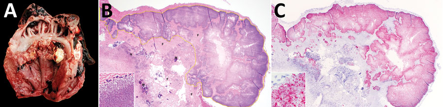
Figure 2. Lesions associated with swine vegetative endocarditis, United States, 2015–2020. A) Macroscopic findings of vegetative growth on the left atrioventricular heart valve leaflets. B) Histopathologic findings of inflammation characterized by necrotic leukocytes (N), fibrin (F), mineralization (M), and myriad bacterial colonization (yellow outline) along the surface of the heart valve (hematoxylin and eosin staining); original magnification ×40. Higher magnification image (inset) shows cocci bacteria in clusters and long chains; original magnification ×1,000. C) Streptococcus gallolyticus directly detected (red) on the surface of the heart valve by RNA in situ hybridization with a probe targeting the helix-hairpin helix domain–containing protein, ComEC/Rec2, and DNA pol III subunit delta genes specific to S. gallolyticus; original magnification ×40. Higher magnification image (inset) shows the bacteria labeled by the in situ hybridization probe; original magnification ×1,000.

Main Article

Page created: November 10, 2021
Page updated: December 20, 2021
Page reviewed: December 20, 2021
The conclusions, findings, and opinions expressed by authors contributing to this journal do not necessarily reflect the official position of the U.S. Department of Health and Human Services, the Public Health Service, the Centers for Disease Control and Prevention, or the authors' affiliated institutions. Use of trade names is for identification only and does not imply endorsement by any of the groups named above.
file_external