Volume 28, Number 11—November 2022
Research Letter
TIGIT Monoallelic Nonsense Variant in Patient with Severe COVID-19 Infection, Thailand
Abstract
A heterozygous nonsense variant in the TIGIT gene was identified in a patient in Thailand who had severe COVID-19, resulting in lower TIGIT expression in T cells. The patient’s T cells produced higher levels of cytokines upon stimulation. This mutation causes less-controlled immune responses, which might contribute to COVID-19 severity.
To investigate SARS-CoV-2 genomic variants, we recruited 46 COVID-19 patients from King Chulalongkorn Memorial Hospital in Bangkok, Thailand, in January 2020. Recruited patients were 16–79 years of age and had moderate to severe COVID-19 symptoms according to World Health Organization interim guidelines (https://apps.who.int/iris/bitstream/handle/10665/331446/WHO-2019-nCoV-clinical-2020.4-eng.pdf). We performed whole-exome sequencing on peripheral blood samples as described (1). The institutional review board of the Faculty of Medicine, Chulalongkorn University, Bangkok, approved this study (COA no. 738/2020).
We filtered variants by using the following criteria. Variants had to pass the quality standards, have read depth >10, and be from the coding regions or canonical splice sites of 1,810 immune-related genes, including immune checkpoint genes (2). Variants also had to have <1% allele frequency in the Genome Aggregation Database (gnomAD, https://gnomad.broadinstitute.org), Exome Variant Server (University of Washington, https://evs.gs.washington.edu/EVS), 1000 Genomes Project Consortium (https://www.genome.gov), dbSNPs (https://www.ncbi.nlm.nih.gov/projects/SNP), and Thai Reference Exome (T-Rex) database (3). We called candidate variants novel pathogenic variants when they were not previously identified in patients in the literature.
In our patient cohort, exome sequencing identified no variants in type I interferon genes, which previously have been commonly observed in patients with severe COVID-19 (4). Of note, we identified a heterozygous nonsense variant (rs1386709957) in the T-cell immunoglobulin and ITIM domain (TIGIT) gene in 1 patient (Appendix Figure 1). We did not identify this nonsense variant among 3,742 persons in the T-Rex database but did observe it in 1 of 31,390 alleles in the gnomAD database, in an allele from a female patient from East Asia. This variant truncates the 245-amino acid residue proteins at residue 56 and is classified as a pathogenic variant American College of Medical Genetics guidelines (https://www.acmg.net).
Figure
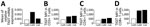
Figure. Results of rRT-PCR assay and flow cytometry of TIGIT nonsense variant in a patient with severe COVID-19 infection, Thailand. Co45 is the patient with TIGITnonsense variant;...
We investigated TIGIT gene expression in T cells of the patient from our study (Co45), a 43-year-old man, and compared it with 2 other sex- and age-matched patients who had severe COVID-19 (Co6 and Co84) (Appendix). We collected peripheral blood mononuclear cells (PBMCs) from each of the patients 1 month after they recovered. We used RNA extracted from PBMCs for real-time reverse transcription PCR and found patient Co45 had the lowest TIGIT mRNA level (Figure, panel A). Because TIGIT is mainly expressed in T cells, we used flow cytometry to measure the mean fluorescence intensity of TIGIT expressed in the cytoplasmic domain (CD) T cells. Patient Co45 had lower TIGIT gene expression in all CD3+, CD4+, and CD8+ T cells than the other 2 patients, most remarkably in the CD8+ T cells (Figure, panels B–D). The percentages of CD3+, CD4+, and CD8+ T cells in patient Co45 were comparable those in the other 2 patients (Appendix Figure 2, panel A), demonstrating that the truncated TIGIT variant reduced TIGIT expression in CD3+, CD4+, and CD8+ T cells.
TIGIT is known to exert immune suppressive functions, such as inhibiting T cell activation, proliferation, and functions that inhibit inflammation and anti-tumor responses. Thus, we investigated the effect of this monoallelic TIGIT variant on T cell functions by examining activation markers and cytokine-secreting T cells after stimulation with anti-CD3/CD28 coupled beads for 24 hours. We then assessed activation by using flow cytometry. We found no differences in frequencies of CD69-expressing CD3+, CD4+, and CD8+ T cells among the 3 patients (Appendix Figure 2, panel B); however, patient Co45 had higher interferon gamma (IFNγ), tumor necrosis factor alpha (TNF-α), and interleukin (IL) 2–producing CD3+, CD4+, and CD8+ T cells than the other 2 patients (Appendix Figure 3).
We believe this patient’s heterozygous nonsense TIGIT variant contributed to the increased inflammatory cytokine functions we observed. His serum cytokine levels at acute illness onset did not differ from the other 2 COVID-19 patients (Appendix Figure 4), but some of his cytokine levels, including IL-10, IL-12p70, IL-4, and IL-7, remained high 1 month after recovery. Upregulation of co-inhibitory receptors, including cytotoxic T-lymphocyte–associated protein 4, programmed cell death protein 1, lymphocyte-activation gene 3, and T-cell immunoglobulin mucin-3, including TIGIT, has been reported in COVID-19 patients in other studies (5). These co-inhibitory receptors upregulated after T-cell activation to regulate immune responses and limit immunopathology (6,7). TIGIT can modulate expression of proinflammatory cytokines in acute lymphocytic choriomeningitis virus infection, in which the TIGIT blockage increased TNF-α expression by CD8+ T cells (8). TIGIT -deficient mice displayed increased IFNγ and IL-17+CD4+ T-cell frequencies (9). Similarly, TIGIT knockdown can increase IFNγ expression in human T cells (10). We hypothesize that the nonsense TIGIT variant led to low TIGIT expression and hyperactive T responses in patient Co45 and might have contributed to his severe inflammation and symptoms. Unfortunately, the patient refused follow-up, so we could not perform further investigations to confirm our hypothesis.
In conclusion, we identified a patient with severe COVID-19 and a TIGIT monoallelic nonsense variant. He had lower TIGIT expression in CD3+, CD4+, and CD8+ T cells and produced higher cytokine expression, including IFNγ, TNF-α, and IL-2 upon stimulation. Our findings suggest TIGIT could be involved in COVID-19 severity.
Dr. Sodsai is a researcher in the Center of Excellence in Immunology and Immune-mediated Diseases, Department of Microbiology, Faculty of Medicine, Chulalongkorn University. Her primary research interests focus on cellular immunology.
Acknowledgment
This study was supported by Ratchadapisek Somphot Fund (grant no RA(PO)005/63); Ratchadapisek Somphot Matching Fund, and Health Systems Research Institute (no. 65-040); e-ASIA Joint Research Program (e-ASIA JRP) administered by the National Science and Technology Development Agency; the Center of Excellence in Immunology and Immune-mediated Diseases; the Center of Excellence for Medical Genomics, Medical Genomics Cluster, Department of Pediatrics; the Center of Excellence in Pediatric Infectious Diseases and Vaccines, Faculty of Medicine, Chulalongkorn University; the Excellence Center for Genomics and Precision Medicine; the Emerging Infectious Diseases Clinical Centre, King Chulalongkorn Memorial Hospital; and The Thai Red Cross Society. Biospecimen collection was supported by Biobank, and the Faculty of Medicine, Chulalongkorn University, Bangkok, Thailand.
References
- Ittiwut R, Sengpanich K, Lauhasurayotin S, Ittiwut C, Shotelersuk V, Sosothikul D, et al. Clinical and molecular characteristics of Thai patients with ELANE-related neutropaenia. J Clin Pathol. 2022;75:99–103. DOIPubMedGoogle Scholar
- Hu FF, Liu CJ, Liu LL, Zhang Q, Guo AY. Expression profile of immune checkpoint genes and their roles in predicting immunotherapy response. Brief Bioinform. 2021;22:bbaa176.
- Shotelersuk V, Wichadakul D, Ngamphiw C, Srichomthong C, Phokaew C, Wilantho A, et al. The Thai reference exome (T-REx) variant database. Clin Genet. 2021;100:703–12. DOIPubMedGoogle Scholar
- Gray PE, Bartlett AW, Tangye SG. Severe COVID-19 represents an undiagnosed primary immunodeficiency in a high proportion of infected individuals. Clin Transl Immunology. 2022;11:
e1365 . DOIPubMedGoogle Scholar - Barnova M, Bobcakova A, Urdova V, Kosturiak R, Kapustova L, Dobrota D, et al. Inhibitory immune checkpoint molecules and exhaustion of T cells in COVID-19. Physiol Res. 2021;70(S2):S227–47. DOIPubMedGoogle Scholar
- Anderson AC, Joller N, Kuchroo VK. Lag-3, Tim-3, and TIGIT: co-inhibitory receptors with specialized functions in immune regulation. Immunity. 2016;44:989–1004. DOIPubMedGoogle Scholar
- Harjunpää H, Guillerey C. TIGIT as an emerging immune checkpoint. Clin Exp Immunol. 2020;200:108–19. DOIPubMedGoogle Scholar
- Schorer M, Rakebrandt N, Lambert K, Hunziker A, Pallmer K, Oxenius A, et al. TIGIT limits immune pathology during viral infections. Nat Commun. 2020;11:1288. DOIPubMedGoogle Scholar
- Joller N, Hafler JP, Brynedal B, Kassam N, Spoerl S, Levin SD, et al. Cutting edge: TIGIT has T cell-intrinsic inhibitory functions. J Immunol. 2011;186:1338–42. DOIPubMedGoogle Scholar
- Lozano E, Dominguez-Villar M, Kuchroo V, Hafler DA. The TIGIT/CD226 axis regulates human T cell function. J Immunol. 2012;188:3869–75. DOIPubMedGoogle Scholar
Figure
Cite This ArticleOriginal Publication Date: October 03, 2022
1These first authors contributed equally to this article.
2These authors were co–principal investigators.
Table of Contents – Volume 28, Number 11—November 2022
| EID Search Options |
|---|
|
|
|
|
|
|
Please use the form below to submit correspondence to the authors or contact them at the following address:
Vorasuk Shotelersuk, Center of Excellence for Medical Genomics, Medical Genomics Cluster, Department of Pediatrics, Faculty of Medicine, Chulalongkorn University, Rama 4 Rd, Bangkok 10330, Thailand
Top