Skip directly to site content Skip directly to page options Skip directly to A-Z link Skip directly to A-Z link Skip directly to A-Z link
Volume 28, Number 2—February 2022
Dispatch

Tonate Virus and Fetal Abnormalities, French Guiana, 2019

Veronique LambertComments to Author , Antoine Enfissi, Mathilde Lefebvre, Leo Pomar, Sobhi Kedous, Fabien Guimiot, Gabriel Carles, Anne Lavergne, Dominique Rousset, and Najeh Hcini
Author affiliations: West French Guiana Hospital Center, Saint-Laurent-du-Maroni, French Guiana (V. Lambert, L. Pomar, S. Kedous, G. Carles, N. Hcini); Institut Pasteur Arbovirus National Reference Center, Cayenne, French Guiana (A. Enfissi, A. Lavergne, D. Rousset); Centre Hospitalo-Universitaire Robert Debre, Paris, France (M. Lefebvre, F. Guimiot); University of French Guiana, Cayenne, French Guiana (N. Hcini)

Main Article

Figure 2

Pathologic findings including results of external examination, histological features of central nervous system, and immunohistochemical staining in a fetus from a woman in French Guiana who was found to be infected with Tonate virus. A) External examination of the body showing subcutaneous edema, microcephaly, craniofacial malformations (short forehead, flat midface), and severe arthrogryposis with upper and lower limb malformations with joint contractures. B) Histologic view of brain section stained in hematoxylin and eosin, displaying lateral ventricle enlargement (asterisk), meningeal hemorrhage (long arrow), diffuse calcifications (short arrows), and nodular heterotopia (arrowheads). Scale bar = 3 mm. C) Spinal cord section showing an abnormally shaped and atrophic spinal cord with the presence of siderophages (sign of premortem meningeal hemorrhage, arrow). Scale bar = 1 mm. D) CD68 immunohistochemistry demonstrating microglial activation and small clusters of microglia and macrophages in the brain (hematoxylin counterstain). Scale bar = 1 mm. E–G) Immunohistochemistry, using anti-TONV mouse serum, of patient (E), control (F), and negative control (G) brains. Note the strong staining of many positive cells in the patient (arrows and inset in panel E), compared to the control brain, where a moderately diffuse background signal is shown but without strong positive cells such as in the patient. In the negative control (without anti-TONV mouse serum antibody), there is a very slight staining (arrowheads and inset in panel G) in the same cells compared with those in the patient, indicating the background signal (color trapping) in these cells. Scale bars = 300 μm; insets in panels E and G = 20 μm.

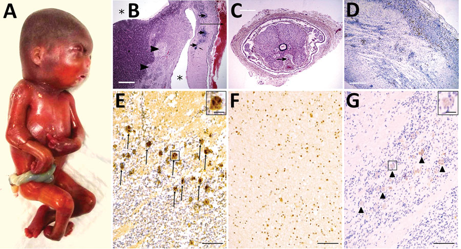
Figure 2. Pathologic findings including results of external examination, histological features of central nervous system, and immunohistochemical staining in a fetus from a woman in French Guiana who was found to be infected with Tonate virus. A) External examination of the body showing subcutaneous edema, microcephaly, craniofacial malformations (short forehead, flat midface), and severe arthrogryposis with upper and lower limb malformations with joint contractures. B) Histologic view of brain section stained in hematoxylin and eosin, displaying lateral ventricle enlargement (asterisk), meningeal hemorrhage (long arrow), diffuse calcifications (short arrows), and nodular heterotopia (arrowheads). Scale bar = 3 mm. C) Spinal cord section showing an abnormally shaped and atrophic spinal cord with the presence of siderophages (sign of premortem meningeal hemorrhage, arrow). Scale bar = 1 mm. D) CD68 immunohistochemistry demonstrating microglial activation and small clusters of microglia and macrophages in the brain (hematoxylin counterstain). Scale bar = 1 mm. E–G) Immunohistochemistry, using anti-TONV mouse serum, of patient (E), control (F), and negative control (G) brains. Note the strong staining of many positive cells in the patient (arrows and inset in panel E), compared to the control brain, where a moderately diffuse background signal is shown but without strong positive cells such as in the patient. In the negative control (without anti-TONV mouse serum antibody), there is a very slight staining (arrowheads and inset in panel G) in the same cells compared with those in the patient, indicating the background signal (color trapping) in these cells. Scale bars = 300 μm; insets in panels E and G = 20 μm.

Main Article

Page created: December 14, 2021
Page updated: January 22, 2022
Page reviewed: January 22, 2022
The conclusions, findings, and opinions expressed by authors contributing to this journal do not necessarily reflect the official position of the U.S. Department of Health and Human Services, the Public Health Service, the Centers for Disease Control and Prevention, or the authors' affiliated institutions. Use of trade names is for identification only and does not imply endorsement by any of the groups named above.
file_external