Skip directly to site content Skip directly to page options Skip directly to A-Z link Skip directly to A-Z link Skip directly to A-Z link
Volume 28, Number 3—March 2022
Synopsis

Detection of SARS-CoV-2 in Neonatal Autopsy Tissues and Placenta

Sarah Reagan-Steiner1Comments to Author , Julu Bhatnagar1, Roosecelis B. Martines, Nicholas S. Milligan, Carly Gisondo, Frank B. Williams, Elizabeth Lee, Lindsey Estetter, Hannah Bullock, Cynthia S. Goldsmith, Pamela Fair, Julie Hand, Gillian Richardson, Kate R. Woodworth, Titilope Oduyebo, Romeo R. Galang, Rebecca Phillips, Elizaveta Belyaeva, Xiao-Ming Yin, Dana Meaney-Delman, Timothy M. Uyeki, Drucilla J. Roberts, and Sherif R. Zaki
Author affiliations: Centers for Disease Control and Prevention, Atlanta, Georgia, USA (S. Reagan-Steiner, J. Bhatnagar, R.B. Martines, E. Lee, L. Estetter, H. Bullock, C.S. Goldsmith, P. Fair, K.R. Woodworth, T. Oduyebo, R.R. Galang, D. Meaney-Delman, T.M. Uyeki, S.R. Zaki); Tulane University School of Medicine, New Orleans, Louisiana, USA (N.S. Milligan, E. Belyaeva, X.-M. Yin); Oschner Health, New Orleans (C. Gisondo, F.B. Williams, R. Phillips); Oak Ridge Institute for Science and Education, Oak Ridge, Tennessee, USA (E. Lee); Synergy America, Inc., Duluth, Georgia, USA (L. Estetter, H. Bullock); Louisiana Department of Health, Baton Rouge, Louisiana, USA (J. Hand, G. Richardson); Massachusetts General Hospital, Boston, Massachusetts, USA (D.J. Roberts)

Main Article

Figure 1

Pulmonary histopathologic, immunohistochemical (IHC), in situ hybridization, and ultrastructural findings in tissues from a neonate in the United States with severe acute respiratory syndrome coronavirus 2 (SARS-CoV-2). A) Lower magnification of the lung showing diffuse alveolar damage, characterized by type II pneumocyte hyperplasia (arrowhead), hyaline membrane (arrow), and interstitial mononuclear infiltrate. Original magnification ×20. B) Extensive intra-alveolar immunostaining by spike protein SARS-CoV-2 IHC assay. Original magnification ×40. C) Double-stain IHC assay showing rare macrophages with SARS-CoV-2/CD-163–positive immunostaining. Red, SARS-CoV-2; brown, CD-163 antibody (arrow). Original magnification ×63. D) Extensive staining of SARS-CoV-2 genomic RNA in pneumocytes by nucleocapsid gene in situ hybridization assay. Original magnification ×10. E) Electron microscopy (EM) image of a pneumocyte containing accumulations of intracellular viral particles. Scale bar indicates 200 nm; viral particles were on average 65 nm in diameter, smaller than commonly observed because of shrinkage during processing. F) Immunostaining of tracheal epithelial cells (arrowhead) and submucosal glands (arrow) by SARS-CoV-2 nucleocapsid IHC assay. Original magnification ×20. G) EM image of a ciliated epithelial cell with extracellular viral particles (arrow) associated with the cilia. Scale bar indicates 200 nm. EM images were collected from 4-μm sections of formalin-fixed, paraffin-embedded tissues affixed to glass slides that were embedded for EM.

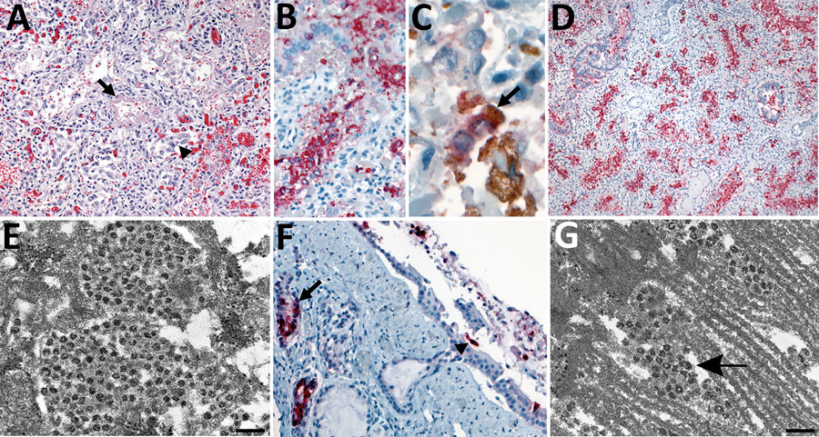
Figure 1. Pulmonary histopathologic, immunohistochemical (IHC), in situ hybridization, and ultrastructural findings in tissues from a neonate in the United States with severe acute respiratory syndrome coronavirus 2 (SARS-CoV-2). A) Lower magnification of the lung showing diffuse alveolar damage, characterized by type II pneumocyte hyperplasia (arrowhead), hyaline membrane (arrow), and interstitial mononuclear infiltrate. Original magnification ×20. B) Extensive intra-alveolar immunostaining by spike protein SARS-CoV-2 IHC assay. Original magnification ×40. C) Double-stain IHC assay showing rare macrophages with SARS-CoV-2/CD-163–positive immunostaining. Red, SARS-CoV-2; brown, CD-163 antibody (arrow). Original magnification ×63. D) Extensive staining of SARS-CoV-2 genomic RNA in pneumocytes by nucleocapsid gene in situ hybridization assay. Original magnification ×10. E) Electron microscopy (EM) image of a pneumocyte containing accumulations of intracellular viral particles. Scale bar indicates 200 nm; viral particles were on average 65 nm in diameter, smaller than commonly observed because of shrinkage during processing. F) Immunostaining of tracheal epithelial cells (arrowhead) and submucosal glands (arrow) by SARS-CoV-2 nucleocapsid IHC assay. Original magnification ×20. G) EM image of a ciliated epithelial cell with extracellular viral particles (arrow) associated with the cilia. Scale bar indicates 200 nm. EM images were collected from 4-μm sections of formalin-fixed, paraffin-embedded tissues affixed to glass slides that were embedded for EM.

Main Article

1These authors contributed equally to this article.

Page created: December 02, 2021
Page updated: February 21, 2022
Page reviewed: February 21, 2022
The conclusions, findings, and opinions expressed by authors contributing to this journal do not necessarily reflect the official position of the U.S. Department of Health and Human Services, the Public Health Service, the Centers for Disease Control and Prevention, or the authors' affiliated institutions. Use of trade names is for identification only and does not imply endorsement by any of the groups named above.
file_external