Skip directly to site content Skip directly to page options Skip directly to A-Z link Skip directly to A-Z link Skip directly to A-Z link
Volume 28, Number 4—April 2022
Dispatch

Rigidoporus corticola Colonization and Invasive Fungal Disease in Immunocompromised Patients, United States

Alvaro C. LagaComments to Author , Jessica W. Crothers, Connie F. Cañete-Gibas, Nathan P. Wiederhold, and Isaac H. Solomon
Author affiliations: Brigham and Women’s Hospital and Harvard Medical School, Boston, Massachusetts, USA (A.C. Laga, I.H. Solomon); University of Vermont Medical Center, Burlington, Vermont, USA (J.W. Crothers); University of Texas Health San Antonio, San Antonio, Texas, USA (C.F. Cañete-Gibas, N.P. Wiederhold)

Main Article

Figure 2

Radiologic and pathologic findings in a 61-year-old immunocompromised man with a history of lung adenocarcinoma and a new 2.4 cm right upper lobe mass determined to be a pulmonary fungus ball caused by Rigidoporus corticola (Oxyporus corticola) infection, United States. A) Chest computed tomography scan. Arrow indicates a 2.4 × 2.2 cm ovoid pulmonary mass in the right upper lobe. B) Hematoxylin and eosin–stained histologic sections of the resected mass. Black arrow indicates a cavitary lesion with a necrotic center. White arrows indicate peripheral fibrous capsule. Original magnification ×100. C, D) Gomori methenamine silver–stained histologic sections. C) Arrows indicate numerous hyphae within the cavity, but no evident invasion into blood vessels or surrounding tissue. Original magnification ×40. D) Arrow indicates septate thin hyphae evident in the center of the cavity. Original magnification ×600.

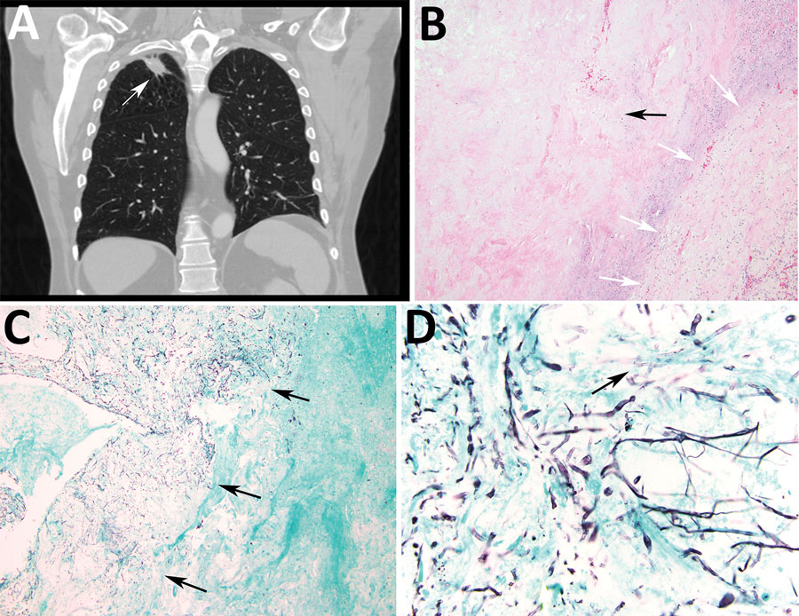
Figure 2. Radiologic and pathologic findings in a 61-year-old immunocompromised man with a history of lung adenocarcinoma and a new 2.4 cm right upper lobe mass determined to be a pulmonary fungus ball caused by Rigidoporus corticola (Oxyporus corticola) infection, United States. A) Chest computed tomography scan. Arrow indicates a 2.4 × 2.2 cm ovoid pulmonary mass in the right upper lobe. B) Hematoxylin and eosin–stained histologic sections of the resected mass. Black arrow indicates a cavitary lesion with a necrotic center. White arrows indicate peripheral fibrous capsule. Original magnification ×100. C, D) Gomori methenamine silver–stained histologic sections. C) Arrows indicate numerous hyphae within the cavity, but no evident invasion into blood vessels or surrounding tissue. Original magnification ×40. D) Arrow indicates septate thin hyphae evident in the center of the cavity. Original magnification ×600.

Main Article

Page created: February 24, 2022
Page updated: March 19, 2022
Page reviewed: March 19, 2022
The conclusions, findings, and opinions expressed by authors contributing to this journal do not necessarily reflect the official position of the U.S. Department of Health and Human Services, the Public Health Service, the Centers for Disease Control and Prevention, or the authors' affiliated institutions. Use of trade names is for identification only and does not imply endorsement by any of the groups named above.
file_external