Volume 28, Number 6—June 2022
Research Letter
Lyme Disease, Anaplasmosis, and Babesiosis, Atlantic Canada
Ziyad O. Allehebi, Farhan M. Khan, Mark Robbins, Elizabeth Simms, Richard Xiang, Allam Shawwa, L. Robbin Lindsay, Antonia Dibernardo, Clarice d’Entremont, Alex Crowell, Jason J. LeBlanc, and David J. Haldane
Figure
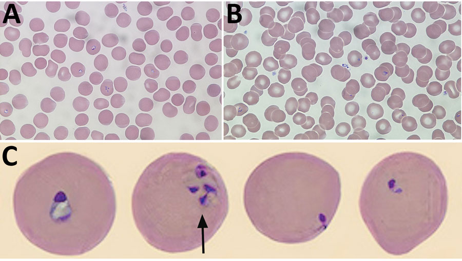
Figure. Babesia microti detected on Wright-stained peripheral blood smears from a 58-year-old man, southwest Nova Scotia, Canada, July 2021. Some typical features of B. microti infection include multiple ring forms present in erythrocytes (A), extracellular ring forms (B), and ring forms of various shapes and sizes (C), including the pathognomonic finding of merozoites arranged in a tetrad formation resembling a Maltese cross (arrow). Images in panels A and B obtained by using Wright’s stain (original magnification ×100), For panel C, the CellaVision DM96 system (https://www.cellavision.com) and the Cellavision Remote Review Software version 6.0.1 build 7 were used to capture and display cells with abnormalities.
Page created: May 13, 2022
Page updated: May 22, 2022
Page reviewed: May 22, 2022
The conclusions, findings, and opinions expressed by authors contributing to this journal do not necessarily reflect the official position of the U.S. Department of Health and Human Services, the Public Health Service, the Centers for Disease Control and Prevention, or the authors' affiliated institutions. Use of trade names is for identification only and does not imply endorsement by any of the groups named above.