Skip directly to site content Skip directly to page options Skip directly to A-Z link Skip directly to A-Z link Skip directly to A-Z link
Volume 28, Number 7—July 2022
Dispatch

Anncaliia algerae Microsporidiosis Diagnosed by Metagenomic Next-Generation Sequencing, China

Chen Liu1, Qin Chen1, Ping Fu, and Yun-Ying ShiComments to Author 
Author affiliations: West China Hospital of Sichuan University, Chengdu, China (C. Liu, Q. Chen, P. Fu, Y.-Y. Shi); Chengdu 363 Hospital of Southwest Medical University, Chengdu (Q. Chen)

Main Article

Figure 2

Light microscopy and transmission electron microscopy of left biceps branchii muscle biopsy tissue from a 45-year-old man with microsporidiosis caused by Anncaliia algerae, China. A–D) Light microscopy using different stains. A) Periodic acid-Schiff stain. Scale bar indicates 10 µm. Original magnification ×50. B) Gomori methenamine silver stain. Scale bar indicates 10 µm. Original magnification ×63. C) Warthin-Starry stain. Scale bar indicates 10 µm. Original magnification ×63. D) Toluidine blue stain. Scale bar indicates 10 µm. Arrows indicate myocytes replaced by aggregates of 2–3 µm ovoid organisms. Original magnification ×63. E, F) Transmission electron microscopy showing Anncaliia-like microsporidia. Scale bars indicate 500 nm. E) A mature spore with electron-dense exospore, electron-lucent endospore, and a single row of 6 to 8 polar tubule coils (arrows). Original magnification ×8,000. F) Proliferating form of microsporidia showing diplokaryotic nuclei (stars) with vesiculotubular structures extending from the meront cell membrane and aggregating in the host cell cytoplasm (arrows). Original magnification ×3,000.

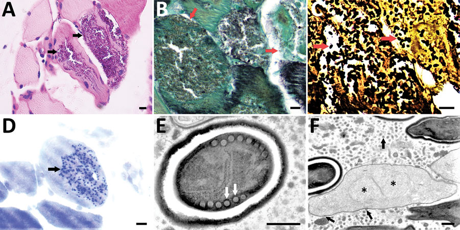
Figure 2. Light microscopy and transmission electron microscopy of left biceps branchii muscle biopsy tissue from a 45-year-old man with microsporidiosis caused by Anncaliia algerae, China. A–D) Light microscopy using different stains. A) Periodic acid-Schiff stain. Scale bar indicates 10 µm. Original magnification ×50. B) Gomori methenamine silver stain. Scale bar indicates 10 µm. Original magnification ×63. C) Warthin-Starry stain. Scale bar indicates 10 µm. Original magnification ×63. D) Toluidine blue stain. Scale bar indicates 10 µm. Arrows indicate myocytes replaced by aggregates of 2–3 µm ovoid organisms. Original magnification ×63. E, F) Transmission electron microscopy showing Anncaliia-like microsporidia. Scale bars indicate 500 nm. E) A mature spore with electron-dense exospore, electron-lucent endospore, and a single row of 6 to 8 polar tubule coils (arrows). Original magnification ×8,000. F) Proliferating form of microsporidia showing diplokaryotic nuclei (stars) with vesiculotubular structures extending from the meront cell membrane and aggregating in the host cell cytoplasm (arrows). Original magnification ×3,000.

Main Article

1These first authors contributed equally to this article.

Page created: May 16, 2022
Page updated: June 18, 2022
Page reviewed: June 18, 2022
The conclusions, findings, and opinions expressed by authors contributing to this journal do not necessarily reflect the official position of the U.S. Department of Health and Human Services, the Public Health Service, the Centers for Disease Control and Prevention, or the authors' affiliated institutions. Use of trade names is for identification only and does not imply endorsement by any of the groups named above.
file_external