Volume 28, Number 8—August 2022
Research Letter
Invasive Streptococcus oralis Expressing Serotype 3 Pneumococcal Capsule, Japan
Abstract
We report 2 adult cases of invasive disease in Japan caused by Streptococcus oralis that expressed the serotype 3 pneumococcal capsule and formed mucoid colonies. Whole-genome sequencing revealed that the identical serotype 3 pneumococcal capsule locus and hyl fragment were recombined into the genomes of 2 distinct S. oralis strains.
Streptococcus oralis is a viridans streptococcus that is divided into 3 subspecies S. oralis subsp. oralis, dentisani, and tigurinus (1). Differentiation between these subspecies and other α-hemolytic streptococci, including S. pneumoniae, remains difficult because they share similar biochemical properties. S. oralis inhabits the oral cavity and can cause severe infections in persons with immunodeficiency (2). Antimicrobial drug resistance and capsule expression studies have demonstrated that gene transfer can occur from oral Streptococcus spp. to S. pneumoniae (3–5). Most oral Streptococcus spp. have a pneumococcus-like capsule locus and produce capsular polysaccharides (6).
We report 2 cases of invasive streptococcal disease in older adults in Japan (Table). Case 1 occurred in a 69-year-old man with gastric cancer; case 2 occurred in a 78-year-old man with bacteremic meningitis who had no underlying disease. Both patients were successfully treated with antimicrobial agents. The bacterial isolates (ASP0312-Sp from case 1 and SP2752 from case 2) contained α-hemolytic bacteria that formed characteristic mucoid colonies on blood agar (Table). Quellung reactions were strongly positive for pool R or pneumococcal serotype 3 antisera (Statens Serum Institut, https://en.ssi.dk), suggesting that the isolates were S. pneumoniae serotype 3. However, both isolates were optochin-resistant and bile-insoluble. Moreover, multilocus sequence typing (MLST) showed that the sequences of all 7 alleles of ASP0312-Sp and 5 alleles of SP2752 differed from those registered in the MLST database (https://pubmlst.org) (Table). For SP2752, the allele numbers were 341 for gdh and 406 for spi. Furthermore, we observed nucleotide differences between ASP0312-Sp and SP2752 in aroE (31 different bp), gdh (34 bp), gki (25 bp), recP (25 bp), spi (14 bp), xpt (47 bp), and ddl (15 bp), which indicated that the strains were distinct. These results suggested that the 2 strains were nonpneumococcal Streptococcus spp.
Figure
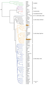
Figure. Phylogenetic analysis of invasive Streptococcus oralisexpressing serotype 3 pneumococcal capsule from 2 adult patients, Japan. Asterisks and orange shading indicate genomes from isolates ASP0312-Sp and SP2752...
For species identification, we performed phylogenetic analyses of whole-genome sequences (Appendix). Homologous core gene clustering showed that ASP0312-Sp and SP2752 belonged to the S. oralis clade (Figure); they were distant from one another, which was consistent with the MLST results.
To investigate recombination events, we compared the sequences surrounding the capsule loci of ASP0312-Sp and SP2752 with those of S. oralis subsp. tigurinus osk_001 and S. pneumoniae serotype 3 OXC141 (Appendix Figure). For ASP0312-Sp, the sequence corresponding to the downstream region of nsik up to the 5′ terminus of the gene encoding the cell wall binding repeat protein in osk_001 was replaced by a fragment of ≈30 kb from pneumococcus. For SP2752, the sequence encoding an ATPase up to the 5′ terminus of the gene encoding the cell wall binding repeat protein in osk_001 was replaced by a fragment of ≈16 kb from pneumococcus. The capsule sequences of ASP0312-Sp and SP2752 were 100% identical to the corresponding sequences located from 303730 to 312820 bp in HU-OH (GenBank accession no. AP018937.1), a serotype 3 pneumococcal strain that was isolated in Japan (7).
We performed homology searches of 36 known pneumococcal virulence genes because multifragment recombination has been demonstrated during the capsular transformation process in pneumococcal populations (8). In ASP0312-Sp and SP2752, the hyl gene, which encodes hyaluronate lyase (9), was located distantly from the capsule locus and shared 96% identity with that of S. pneumoniae. We did not detect homologs of the other 35 genes for either isolate.
A recent study reported that acapsular pneumococcus became virulent after transformation with the capsule gene from SK95, which is an oral S. mitis strain (5). This previous study demonstrated a cross-species transformation from a commensal streptococcal species to pneumococcus (5). Our results complement this report, although the direction of transformation in our study was reversed. Our analyses of 2 human patients with invasive disease caused by S. oralis provided evidence of cross-species gene transfer from pneumococcus to a commensal streptococcal species. Acquisition of capsule and hyl genes might have increased pathogenicity (9,10) and contributed to progression of invasive disease in these 2 cases.
In conclusion, because of discrepancies between phenotypic and biochemical analyses, we used MLST and whole-genome sequencing to identify streptococcal species in these 2 patients. Our study indicates a potential pitfall for identifying and serotyping pneumococci that can occur if the bacteria are not isolated. Thus, when α-hemolytic streptococci are isolated from a sterile site, clinicians should request molecular analyses to identify the causative species, regardless of the mucoid phenotype.
Dr. Chang is a microbiologist at the National Institute of Infectious Diseases, Tokyo, Japan. Her research interests focus on the microbiology of Streptococcus pneumoniae.
Acknowledgments
We thank Moon H. Nahm for his valuable comments.
This work was supported by grants from the Japan Agency for Medical Research and Development (AMED; no. JP20fk0108139) and Ministry of Health, Labour and Welfare (no. JPMH19HA1005).
References
- Jensen A, Scholz CFP, Kilian M. Re-evaluation of the taxonomy of the Mitis group of the genus Streptococcus based on whole genome phylogenetic analyses, and proposed reclassification of Streptococcus dentisani as Streptococcus oralis subsp. dentisani comb. nov., Streptococcus tigurinus as Streptococcus oralis subsp. tigurinus comb. nov., and Streptococcus oligofermentans as a later synonym of Streptococcus cristatus. Int J Syst Evol Microbiol. 2016;66:4803–20. DOIPubMedGoogle Scholar
- Nakamura Y, Uemura T, Kawata Y, Hirose B, Yamauchi R, Shimohama S. Streptococcus oralis meningitis with gingival bleeding in a patient: a case report and review of the literature. Intern Med. 2021;60:789–93. DOIPubMedGoogle Scholar
- Bryskier A. Viridans group streptococci: a reservoir of resistant bacteria in oral cavities. Clin Microbiol Infect. 2002;8:65–9. DOIPubMedGoogle Scholar
- Ganaie F, Saad JS, McGee L, van Tonder AJ, Bentley SD, Lo SW, et al. A new pneumococcal capsule type, 10D, is the 100th serotype and has a large cps fragment from an oral Streptococcus. MBio. 2020;11:e00937–20. DOIPubMedGoogle Scholar
- Nahm MH, Brissac T, Kilian M, Vlach J, Orihuela CJ, Saad JS, et al. Pneumococci can become virulent by acquiring a new capsule from oral streptococci. J Infect Dis. 2020;222:372–80. DOIPubMedGoogle Scholar
- Skov Sørensen UB, Yao K, Yang Y, Tettelin H, Kilian M. Capsular polysaccharide expression in commensal Streptococcus species: genetic and antigenic similarities to Streptococcus pneumoniae. MBio. 2016;7:e01844–16. DOIPubMedGoogle Scholar
- Nagaoka K, Konno S, Murase K, Kikuchi T, Morinaga Y, Yanagihara K, et al. Complete genome sequences of Streptococcus pneumoniae strains HU-OH (serotype 3, sequence type 183 [ST183]), NU83127 (serotype 4, ST246), and ATCC 49619 (serotype 19F, ST1203). Microbiol Resour Announc. 2019;8:e01504–18. DOIPubMedGoogle Scholar
- Golubchik T, Brueggemann AB, Street T, Gertz RE Jr, Spencer CCA, Ho T, et al. Pneumococcal genome sequencing tracks a vaccine escape variant formed through a multi-fragment recombination event. Nat Genet. 2012;44:352–5. DOIPubMedGoogle Scholar
- Suits MDL, Pluvinage B, Law A, Liu Y, Palma AS, Chai W, et al. Conformational analysis of the Streptococcus pneumoniae hyaluronate lyase and characterization of its hyaluronan-specific carbohydrate-binding module. J Biol Chem. 2014;289:27264–77. DOIPubMedGoogle Scholar
- Kadioglu A, Weiser JN, Paton JC, Andrew PW. The role of Streptococcus pneumoniae virulence factors in host respiratory colonization and disease. Nat Rev Microbiol. 2008;6:288–301. DOIPubMedGoogle Scholar
Figure
Table
Cite This ArticleOriginal Publication Date: July 08, 2022
1Current affiliation: Matsue Red Cross Hospital, Shimane, Japan.
Table of Contents – Volume 28, Number 8—August 2022
| EID Search Options |
|---|
|
|
|
|
|
|
Please use the form below to submit correspondence to the authors or contact them at the following address:
Bin Chang, Department of Bacteriology I, National Institute of Infectious Diseases, 1-23-1, Toyama, Shinjuku-ku, Tokyo 162-8640, Japan
Top