Skip directly to site content Skip directly to page options Skip directly to A-Z link Skip directly to A-Z link Skip directly to A-Z link
Volume 28, Number 9—September 2022
Research Letter

Trichodysplasia Spinulosa Polyomavirus Endothelial Infection, California, USA

Lauren Lawrence1Comments to Author , Aihui Wang, Gregory Charville, Angus Toland, Benjamin Pinsky, Yasodha Natkunam, Sheren Younes, Henning Stehr, and Dita Gratzinger
Author affiliations: Stanford Healthcare, Palo Alto, California, USA (L. Lawrence, A. Toland); Stanford University School of Medicine, Stanford, California, USA (A. Wang, G. Charville, B. Pinsky, Y. Natkunam, S. Younes, H. Stehr, D. Gratzinger)

Main Article

Figure

Tissue samples from 3 patients with trichodysplasia spinulosa polyomavirus endothelial infection, California, USA. We performed hematoxylin and eosin (H&E) staining and RNAScope in situ hybridization (ISH) to detect trichodysplasia spinulosa polyomavirus (TSPyV) in formalin-fixed, paraffin-embedded tissue specimens. Bright red, granular staining in endothelium indicates TSPyV RNA. A) Case 1, H&E staining, original magnification ×10; B) case 1, TSPyV ISH, original magnification ×10; C) case 1, TSPyV ISH, original magnification ×40; D) case 2, H&E staining, original magnification ×5; E) case 2, TSPyV ISH, original magnification ×10; F) case 2, TSPyV ISH, original magnification ×40; G) case 3, H&E staining, original magnification ×5; H) case 3, TSPyV ISH, original magnification ×10; I) case 3, TSPyV ISH, original magnification ×40; J) biopsy from patient with cutaneous trichodysplasia spinulosa (positive control), TSPyV ISH, original magnification ×10; K) cutaneous biopsy from patient with Merkel cell polyomavirus-positive Merkel cell carcinoma (negative control), TSPyV ISH, original magnification ×40; L) renal biopsy from patient with BK polyomavirus nephropathy (negative control), TSPyV ISH, original magnification ×60.

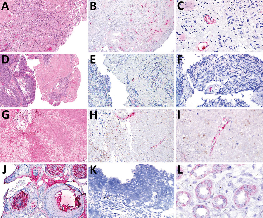
Figure. Tissue samples from 3 patients with trichodysplasia spinulosa polyomavirus endothelial infection, California, USA. We performed hematoxylin and eosin (H&E) staining and RNAScope in situ hybridization (ISH) to detect trichodysplasia spinulosa polyomavirus (TSPyV) in formalin-fixed, paraffin-embedded tissue specimens. Bright red, granular staining in endothelium indicates TSPyV RNA. A) Case 1, H&E staining, original magnification ×10; B) case 1, TSPyV ISH, original magnification ×10; C) case 1, TSPyV ISH, original magnification ×40; D) case 2, H&E staining, original magnification ×5; E) case 2, TSPyV ISH, original magnification ×10; F) case 2, TSPyV ISH, original magnification ×40; G) case 3, H&E staining, original magnification ×5; H) case 3, TSPyV ISH, original magnification ×10; I) case 3, TSPyV ISH, original magnification ×40; J) biopsy from patient with cutaneous trichodysplasia spinulosa (positive control), TSPyV ISH, original magnification ×10; K) cutaneous biopsy from patient with Merkel cell polyomavirus-positive Merkel cell carcinoma (negative control), TSPyV ISH, original magnification ×40; L) renal biopsy from patient with BK polyomavirus nephropathy (negative control), TSPyV ISH, original magnification ×60.

Main Article

1Current affiliation: Guardant Health, Redwood City, California, USA.

Page created: July 07, 2022
Page updated: August 19, 2022
Page reviewed: August 19, 2022
The conclusions, findings, and opinions expressed by authors contributing to this journal do not necessarily reflect the official position of the U.S. Department of Health and Human Services, the Public Health Service, the Centers for Disease Control and Prevention, or the authors' affiliated institutions. Use of trade names is for identification only and does not imply endorsement by any of the groups named above.
file_external