Volume 29, Number 2—February 2023
Research Letter
Orthopoxvirus Infections in Rodents, Nigeria, 2018–2019
Abstract
To investigate animal reservoirs of monkeypox virus in Nigeria, we sampled 240 rodents during 2018–2019. Molecular (real-time PCR) and serologic (IgM) evidence indicated orthopoxvirus infections, but presence of monkeypox virus was not confirmed. These results can be used to develop public health interventions to reduce human infection with orthopoxviruses.
Resurgence of zoonotic agent monkeypox virus (MPXV); genus Orthopoxvirus) in Nigeria since 2017 calls attention to the need to identify the source of primary transmission at the human–animal interface. Results of previous ecologic investigations of the animal reservoir of MPXV have been inconclusive (1,2). However, molecular and serologic evidence suggest that potential reservoirs are rope squirrels (Funisciurus), sun squirrels (Heliosciurus), African pouched rats (Cricetomys), and dormice (Graphiurus) (3–5). In Nigeria, investigations into the ecology and natural history of MPXV and its reservoir are limited (4). Cases of mpox in humans (formerly monkeypox) in Nigeria were reported in 1971 and 1978 but not again until 2017. During 2017–August 2022, a total of 503 cases of MPXV were confirmed, and evidence of exportation from Nigeria is a public health concern (6,7). In addition to vaccinating humans, another feasible public health intervention would be identifying and avoiding spillover from animals (8).
As a preliminary step in identifying the putative MPXV animal reservoir in Nigeria, we sampled 240 rodents during 2018–2019. Humane capture and sampling followed protocols approved by the National Veterinary Research Institute and the Centers for Disease Control and Prevention Animal Care and Use Committees (AEC/03/53/18).
Figure
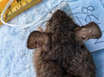
Figure. Rodent N198 with poxlike lesions (arrows) in the ears but negative for orthopoxvirus by quantitative PCR and ELISA, sampled in study of orthopoxvirus infections in rodents, Nigeria, 2018–2019.
We captured and sampled 240 rodents by using a mixture of Tomahawk (https://www.livetrap.com), Sherman (https://www.shermantraps.com), and snap traps in 4 locations: Afi Mountain rain forest, Cross-River state (n = 56); Okomu National Park, Edo state (n = 61); Yenagoa Forest, Bayelsa state (n = 44); and urban and periurban locations associated with recent human cases in Port Harcourt, River state (n = 79). Before collecting blood and euthanizing the rodents, we anesthetized them with inhalant halothane and examined them visually for the presence of poxlike lesions or other signs of illness. If lesions were observed, as seen in animal N198 (Figure), we collected samples from the lesion site. After euthanizing the rodents, we performed necropsies and collected internal organs (e.g., lung, heart, liver, kidney, spleen, skin). For animals not euthanized (as determined for conservation reasons by park officers), we collected only blood and oral and anal swab samples. We extracted DNA by using a MagMAX magnetic processor (ThermoFisher Scientific, https://www.thermofisher.com) and conducted real-time PCR on all available samples by using OPXV-generic and MPXV-specific assays (9). We used ELISA to analyze serum and dried blood spots (5) and considered samples that were reactive at 1:100 and 1:200 dilutions to be positive.
Animal NG114 (Mus baoulei), collected from Okomu National Park, Edo, was positive by generic OPXV PCR of skin, spleen, kidney and by dried blood spot but negative for OPXV antibodies by ELISA. All samples from that animal were negative by MPXV-specific assay. Samples from all other animals were negative by PCR (OPXV-generic and MPXV-specific). Samples from 2 rodents, NG112 (Praomys sp.) and NG173 (Rattus rattus), collected in Okomu National Park on December 12, 2018, and from Port Harcourt, River state (prison environment) on July 5, 2019, were positive by ELISA (OPXV IgG). Khodakevich et al. (4) detected OPXV antibodies or “MPXV–specific” antibodies in rodents (including Praomys) and nonhuman primates in areas where human cases had been reported. A similar investigation in the Democratic Republic of the Congo, conducted in 2012, 2013, and 2015, detected OPXV IgG in 6 rodents, but no rodent was positive by PCR (9). Identifying an unknown OPXV via molecular diagnostics (PCR) from a M. baoulei mouse (NG114) in Okomu and confirmation of ELISA-positive rodents from both urban and rural sites (Port Harcourt and Okomu) indicate the potential role of these animals in the circulation and transmission of OPXVs. The IgG-positive R. rattus rat captured at a prison in Port Harcourt indicates potential OPXV exposure of prisoners and of the local population in urban areas through inconspicuous contact with peridomestic rodents.
Our laboratory analyses showed molecular and serologic evidence of OPXV infections (Table); however, they did not confirm presence of MPXV in the animal samples. Because the ELISA cannot distinguish between different species of OPXVs, we could not determine if the 2 animals with OPXV IgG (NG112 and NG173) were exposed to MPXV or another OPXV circulating in small mammals in Nigeria. Detection of diverse OPXVs in rodents in Nigeria can be used to develop public health interventions to reduce human infection with orthopoxviruses, including MPXV.
Dr. Meseko is a veterinarian and virologist with the National Veterinary Research Institute, Vom, Nigeria. He coordinates the One Health team for field and laboratory investigation of animal reservoirs of MPXV in Nigeria.
Acknowledgments
We thank the Federal and State Ministries of Agriculture, Health and the Environment for their administrative support.
The US Centers for Disease Control and Prevention funded the Monkeypox Animal Reservoir study in Nigeria. The field team (One Health) includes members from the National Veterinary Research Institute, Nigeria Centre for Disease Control, Federal Ministry of Agriculture and Rural Development, and African Field Epidemiology Network.
References
- Khodakevich L, Jezek Z, Messinger D. Monkeypox virus: ecology and public health significance. Bull World Health Organ. 1988;66:747–52.PubMedGoogle Scholar
- Damon IK. Status of human monkeypox: clinical disease, epidemiology and research. Vaccine. 2011;29(Suppl 4):D54–9. DOIPubMedGoogle Scholar
- Reynolds MG, Doty JB, McCollum AM, Olson VA, Nakazawa Y. Monkeypox re-emergence in Africa: a call to expand the concept and practice of One Health. Expert Rev Anti Infect Ther. 2019;17:129–39. DOIPubMedGoogle Scholar
- Khodakevich L, Jezek Z, Kinzanzka K. Isolation of monkeypox virus from wild squirrel infected in nature. Lancet. 1986;1:98–9. DOIPubMedGoogle Scholar
- Doty JB, Malekani JM, Kalemba LN, Stanley WT, Monroe BP, Nakazawa YU, et al. Assessing monkeypox virus prevalence in small mammals at the human-animal interface in the Democratic Republic of the Congo. Viruses. 2017;9:283. DOIPubMedGoogle Scholar
- Alakunle E, Moens U, Nchinda G, Okeke MI. Monkeypox virus in Nigeria: infection biology, epidemiology, and evolution. Viruses. 2020;12:1257. DOIPubMedGoogle Scholar
- Mauldin MR, McCollum AM, Nakazawa YJ, Mandra A, Whitehouse ER, Davidson W, et al. Exportation of monkeypox virus from the African continent. J Infect Dis. 2022;225:1367–76. DOIPubMedGoogle Scholar
- Nolen LD, Osadebe L, Katomba J, Likofata J, Mukadi D, Monroe B, et al. Introduction of monkeypox into a community and household: risk factors and zoonotic reservoirs in the Democratic Republic of the Congo. Am J Trop Med Hyg. 2015;93:410–5. DOIPubMedGoogle Scholar
- Li Y, Olson VA, Laue T, Laker MT, Damon IK. Detection of monkeypox virus with real-time PCR assays. J Clin Virol. 2006;36:194–203. DOIPubMedGoogle Scholar
Figure
Table
Cite This ArticleTable of Contents – Volume 29, Number 2—February 2023
| EID Search Options |
|---|
|
|
|
|
|
|
Please use the form below to submit correspondence to the authors or contact them at the following address:
Clement Meseko, National Veterinary Research Institute, P.B.M. 01 Vom Plateau, Nigeria
Top