Volume 29, Number 4—April 2023
Research Letter
Harbor Porpoise Deaths Associated with Erysipelothrix rhusiopathiae, the Netherlands, 2021
Abstract
In August 2021, a large-scale mortality event affected harbor porpoises (Phocoena phocoena) in the Netherlands. Pathology and ancillary testing of 22 animals indicated that the most likely cause of death was Erysipelothrix rhusiopathiae infection. This zoonotic agent poses a health hazard for cetaceans and possibly for persons handling cetacean carcasses.
Erysipelothrix bacteria cause infections in humans and other species after contact with infected animals or environmental sources (1). Illness ranges from mild to systemic, which can include septicemia and endocarditis. Erysipelothrix can survive for long periods in the environment, including marine ecosystems (1) associated with marine fish, mollusks, and crustaceans. Erysipelothrix infection affects captive and free-ranging crustaceans and is linked to fatal sepsis (2). To our knowledge, reports of large-scale mortality events caused by Erysipelothrix infection in marine mammals are absent from the literature, and Erysipelothrix has not been detected in stranded porpoises along the Netherlands coastline since the start of our harbor porpoise stranding research program in 2008.
At the end of August 2021, a total of 190 dead harbor porpoises (Phocoena phocoena) were found on Dutch Wadden islands; the annual average for stranded harbor porpoises on the entire Dutch coastline is 600. No anthropogenic activities in the southern or central North Sea that could explain this mortality event were reported to the government of the Netherlands in the 4–6 weeks before the event.
Most porpoises were found in an advanced state of decomposition. Twenty-two animals were collected for examination at the Faculty of Veterinary Medicine of Utrecht University (Appendix Table 1). We immediately necropsied 2, and the rest were temporarily frozen pending postmortem investigation and ancillary testing.
Because of advanced decomposition, we could perform only gross pathologic examinations and sampling for ancillary testing, following a standardized international protocol (3). Adult female porpoises were mostly in good to moderate nutritional condition with mild to moderate parasitic infections of various organs and had been reproductively active (Appendix Table 1). Of the 21 stomachs examined (1 was not examined because of gross damage caused by scavengers), none contained marine debris; 10 contained the remains of a few prey, reflecting nonrecent food intake, and the remaining stomachs were empty.
Samples from 3 porpoises with gross changes (mammary gland, lung, spinal cord) were cultured on blood agar (bioTrading, https://biotrading.com) at 37°C for 48 h. Culture results were positive for Erysipelothrix rhusiopathiae. Subsequently, we tested liver samples from 21 animals for E. rhusiopathiae; and 16 were positive (Appendix Table 2). To investigate the relatedness of isolates, genomes of 18 isolates were sequenced by using Illumina NextSeq (https://www.illumina.com) and assembled by using SPAdes version 3.14.1 (4); we included 11 publicly available reference genomes from different E. rhusiopathiae clades (5). A core genome alignment was made with Parsnp verson 1.2 (6) and visualized by using iTol version 4 (7).
Figure
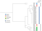
Genomes from this study were phylogenetically positioned between clade 2 E. rhusiopathiae reference genomes and formed 2 distinct clusters showing ≈3,400 single-nucleotide polymorphism (SNP) differences and limited diversity of <6 SNPs within the clusters (Figure). That pattern suggests dissemination of 2 clonal lineages of E. rhusiopathiae, either through exposure to a common source or contact between individuals.
Virology tests on 14 fecal, 15 blood, and 17 spleen samples and metagenomic sequencing with VirCapSeq-VERT (8) revealed no virus sequences of interest. In addition, we tested 20 lung and 20 brain samples for influenza A virus, paramyxoviruses (including morbilliviruses), coronaviruses (including SARS-CoV-2), and herpesviruses. Only 2 brain samples tested positive for P. phocoena alphaherpesvirus (Appendix Table 1), described as an incidental cause of death in porpoises (9). Our results indicate that viruses were an unlikely factor in this mortality event.
We pooled 20 stomach content samples and 21 liver samples in triplicate and analyzed them with a Liquid Chromatograph Triple Quadrupole Mass Spectrometer (LC-MS/MS) (McCrone Associates, https://www.mccrone.com) for domoic acid, saxitoxins, tetrodotoxin, and lipophilic marine toxins. Only saxitoxin was detected; it was in 1 pooled liver sample (estimated concentration 15 μg/kg). Subsequently, we analyzed livers individually, and saxitoxin was not confirmed in any of the individual samples. We therefore conclude that harmful algae were an unlikely factor in this mortality event.
Gross pathologic assessment revealed a moderate to good body condition for most porpoises, but none had recently fed. This finding suggests a subacute cause of death from sudden and excessive disease. No clinically relevant viruses were detected. Phycotoxins were detected in a limited number of porpoises. In contrast, E. rhusiopathiae was isolated from most investigated porpoises. Therefore, we consider E. rhusiopathiae to be the most likely cause of death. Advanced autolysis of the carcasses made detection of distinctive lesions associated with Erysipelothrix infection impossible. The low number of SNPs differing between isolates suggests common exposure, possibly a food source, transmission between porpoises, or both.
Our results draw attention to possibly increased cetacean susceptibility to E. rhusiopathiae, to new or emerging sources of Erysipelothrix in the marine environment, or both. Erysipelothrix remains viable in a carcass up to 12 days in direct sunlight, up to 4 months in putrefied flesh, and up to 9 months in a buried carcass (10). This new emerging source and the long survival time in carcasses demonstrates a need for having only trained personnel handle stranded animals, proper disposal of carcasses, and increased awareness for the potential presence and transmission of this zoonotic bacterium among cetaceans.
Dr. IJsseldijk is assistant professor at the Division of Pathology, Department of Biomolecular Health Sciences, Faculty of Veterinary Medicine, Utrecht University. Her primary research interests focus on marine mammal health and conservation.
Acknowledgments
We thank all volunteers and organizations of the Dutch Stranding Network for their tremendous efforts and work to document stranded porpoises during the event. Necropsies were assisted by Louis van den Boom, Natashja Ennen-Buijs, Olle Juch, Darryl Leydekkers, Immelie Coenen Morales, Jan Mosterd, Eva Schotanus, and Ruby Wagensveld. We also thank Judith van den Brand for her input in the discussions of the ancillary testing. Virology was conducted at Erasmus MC, with special thanks to Marco van de Bildt, Irina Chestakova, Marjan Boter, Babette Weller, Reina Sikkema, and Marion Koopmans. Bacteriology was conducted at the Veterinary Microbiological Diagnostic Center, for which we thank all staff involved. Stomach investigations were conducted at Wageningen Marine Research with the help of Guido Keijl and Eva Schotanus. Algae toxin research was carried out by Wageningen Food Safety Research with the help of Domenique van der Horst.
The research was financed by the Ministry of Agriculture, Nature and Food Quality (project no. 1400012118), for which we are especially grateful for the help of Anne-Marie Svoboda and Sandra van der Graaf.
References
- Wang Q, Chang BJ, Riley TV. Erysipelothrix rhusiopathiae. Vet Microbiol. 2010;140:405–17. DOIPubMedGoogle Scholar
- Ceccolini ME, Wessels M, Macgregor SK, Deaville R, Perkins M, Jepson PD, et al. Systemic Erysipelothrix rhusiopathiae in seven free-ranging delphinids stranded in England and Wales. Dis Aquat Organ. 2021;145:173–84. DOIPubMedGoogle Scholar
- IJsseldijk LL, Brownlow AC, Mazzariol S, editors. Best practice for cetacean post mortem investigation and tissue sampling. Joint ACCOBAMS and ASCOBANS document. Stralsund, Germany: Agreement for the Conservation of Cetaceans of the Baltic Sea, Mediterranean Sea and Contiguous Atlantic Area (ACCOBAMS); Agreement on the Conservation of Small Cetaceans of the Baltic, North East Atlantic, Irish and North Seas (ASCOBANS); 2019 [cited 2022 Nov 7]. https://www.ascobans.org/sites/default/files/document/ascobans_ac25_inf3.2_rev1_best-practice-cetacean-post-mortem-investigation.pdf
- Bankevich A, Nurk S, Antipov D, Gurevich AA, Dvorkin M, Kulikov AS, et al. SPAdes: a new genome assembly algorithm and its applications to single-cell sequencing. J Comput Biol. 2012;19:455–77. DOIPubMedGoogle Scholar
- Forde T, Biek R, Zadoks R, Workentine ML, De Buck J, Kutz S, et al. Genomic analysis of the multi-host pathogen Erysipelothrix rhusiopathiae reveals extensive recombination as well as the existence of three generalist clades with wide geographic distribution. BMC Genomics. 2016;17:461. DOIPubMedGoogle Scholar
- Treangen TJ, Ondov BD, Koren S, Phillippy AM. The Harvest suite for rapid core-genome alignment and visualization of thousands of intraspecific microbial genomes. Genome Biol. 2014;15:524. DOIPubMedGoogle Scholar
- Letunic I, Bork P. Interactive Tree Of Life (iTOL) v4: recent updates and new developments. Nucleic Acids Res. 2019;47(W1):W256–9. DOIPubMedGoogle Scholar
- Briese T, Kapoor A, Mishra N, Jain K, Kumar A, Jabado OJ, et al. Virome capture sequencing enables sensitive viral diagnosis and comprehensive virome analysis. MBio. 2015;6:e01491–15. DOIPubMedGoogle Scholar
- van Elk C, van de Bildt M, van Run P, de Jong A, Getu S, Verjans G, et al. Central nervous system disease and genital disease in harbor porpoises (Phocoena phocoena) are associated with different herpesviruses. Vet Res (Faisalabad). 2016;47:28. DOIPubMedGoogle Scholar
- Brooke CJ, Riley TV. Erysipelothrix rhusiopathiae: bacteriology, epidemiology and clinical manifestations of an occupational pathogen. J Med Microbiol. 1999;48:789–99. DOIPubMedGoogle Scholar
Figure
Cite This ArticleOriginal Publication Date: March 18, 2023
Table of Contents – Volume 29, Number 4—April 2023
| EID Search Options |
|---|
|
|
|
|
|
|
Please use the form below to submit correspondence to the authors or contact them at the following address:
Els M. Broens, Department of Biomolecular Health Sciences, Faculty of Veterinary Medicine, Utrecht University, Yalelaan 1, 3584 TD, Utrecht, the Netherlands
Top