Synopses
Zika virus infection during pregnancy can lead to congenital Zika syndrome. Implementation of screening programs and interpretation of test results can be particularly challenging during ongoing local mosquitoborne transmission. We conducted a retrospective chart review of 2,327 pregnant women screened for Zika virus in Miami–Dade County, Florida, USA, during 2016. Of these, 86 had laboratory evidence of Zika virus infection; we describe 2 infants with probable congenital Zika syndrome. Delays in receipt of laboratory test results (median 42 days) occurred during the first month of local transmission. Odds of screening positive for Zika virus were higher for women without health insurance or who did not speak English. Our findings indicate the increase in screening for Zika virus can overwhelm hospital and public health systems, resulting in delayed receipt of results of screening and confirmatory tests and the potential to miss cases or delay diagnoses.
| EID | Shiu C, Starker R, Kwal J, Bartlett M, Crane A, Greissman S, et al. Zika Virus Testing and Outcomes during Pregnancy, Florida, USA, 2016. Emerg Infect Dis. 2018;24(1):1-8. https://doi.org/10.3201/eid2401.170979 |
|---|---|
| AMA | Shiu C, Starker R, Kwal J, et al. Zika Virus Testing and Outcomes during Pregnancy, Florida, USA, 2016. Emerging Infectious Diseases. 2018;24(1):1-8. doi:10.3201/eid2401.170979. |
| APA | Shiu, C., Starker, R., Kwal, J., Bartlett, M., Crane, A., Greissman, S....Curry, C. L. (2018). Zika Virus Testing and Outcomes during Pregnancy, Florida, USA, 2016. Emerging Infectious Diseases, 24(1), 1-8. https://doi.org/10.3201/eid2401.170979. |
Research
Sensitivity and Specificity of Suspected Case Definition Used during West Africa Ebola Epidemic [PDF - 409 KB - 6 pages]
Rapid early detection and control of Ebola virus disease (EVD) is contingent on accurate case definitions. Using an epidemic surveillance dataset from Guinea, we analyzed an EVD case definition developed by the World Health Organization (WHO) and used in Guinea. We used the surveillance dataset (March–October 2014; n = 2,847 persons) to identify patients who satisfied or did not satisfy case definition criteria. Laboratory confirmation determined cases from noncases, and we calculated sensitivity, specificity and predictive values. The sensitivity of the defintion was 68.9%, and the specificity of the definition was 49.6%. The presence of epidemiologic risk factors (i.e., recent contact with a known or suspected EVD case-patient) had the highest sensitivity (74.7%), and unexplained deaths had the highest specificity (92.8%). Results for case definition analyses were statistically significant (p<0.05 by χ2 test). Multiple components of the EVD case definition used in Guinea contributed to improved overall sensitivity and specificity.
| EID | Hsu CH, Champaloux SW, Keïta S, Martel L, Bilivogui P, Knust B, et al. Sensitivity and Specificity of Suspected Case Definition Used during West Africa Ebola Epidemic. Emerg Infect Dis. 2018;24(1):9-14. https://doi.org/10.3201/eid2401.161678 |
|---|---|
| AMA | Hsu CH, Champaloux SW, Keïta S, et al. Sensitivity and Specificity of Suspected Case Definition Used during West Africa Ebola Epidemic. Emerging Infectious Diseases. 2018;24(1):9-14. doi:10.3201/eid2401.161678. |
| APA | Hsu, C. H., Champaloux, S. W., Keïta, S., Martel, L., Bilivogui, P., Knust, B....McCollum, A. M. (2018). Sensitivity and Specificity of Suspected Case Definition Used during West Africa Ebola Epidemic. Emerging Infectious Diseases, 24(1), 9-14. https://doi.org/10.3201/eid2401.161678. |
Nipah Virus Contamination of Hospital Surfaces during Outbreaks, Bangladesh, 2013–2014 [PDF - 596 KB - 7 pages]
Nipah virus (NiV) has been transmitted from patient to caregivers in Bangladesh presumably through oral secretions. We aimed to detect whether NiV-infected patients contaminate hospital surfaces with the virus. During December 2013–April 2014, we collected 1 swab sample from 5 surfaces near NiV-infected patients and tested surface and oral swab samples by real-time reverse transcription PCR for NiV RNA. We identified 16 Nipah patients; 12 cases were laboratory-confirmed and 4 probable. Of the 12 laboratory-confirmed cases, 10 showed NiV RNA in oral swab specimens. We obtained surface swab samples for 6 Nipah patients; 5 had evidence of NiV RNA on >1 surface: 4 patients contaminated towels, 3 bed sheets, and 1 the bed rail. Patients with NiV RNA in oral swab samples were significantly more likely than other Nipah patients to die. To reduce the risk for fomite transmission of NiV, infection control should target hospital surfaces.
| EID | Hassan M, Sazzad H, Luby SP, Sturm-Ramirez K, Bhuiyan M, Rahman M, et al. Nipah Virus Contamination of Hospital Surfaces during Outbreaks, Bangladesh, 2013–2014. Emerg Infect Dis. 2018;24(1):15-21. https://doi.org/10.3201/eid2401.161758 |
|---|---|
| AMA | Hassan M, Sazzad H, Luby SP, et al. Nipah Virus Contamination of Hospital Surfaces during Outbreaks, Bangladesh, 2013–2014. Emerging Infectious Diseases. 2018;24(1):15-21. doi:10.3201/eid2401.161758. |
| APA | Hassan, M., Sazzad, H., Luby, S. P., Sturm-Ramirez, K., Bhuiyan, M., Rahman, M....Gurley, E. S. (2018). Nipah Virus Contamination of Hospital Surfaces during Outbreaks, Bangladesh, 2013–2014. Emerging Infectious Diseases, 24(1), 15-21. https://doi.org/10.3201/eid2401.161758. |
Detection and Circulation of a Novel Rabbit Hemorrhagic Disease Virus in Australia [PDF - 1.83 MB - 9 pages]
The highly virulent rabbit hemorrhagic disease virus (RHDV) has been widely used in Australia and New Zealand since the mid-1990s to control wild rabbits, an invasive vertebrate pest in these countries. In January 2014, an exotic RHDV was detected in Australia, and 8 additional outbreaks were reported in both domestic and wild rabbits in the 15 months following its detection. Full-length genomic analysis revealed that this virus is a recombinant containing an RHDVa capsid gene and nonstructural genes most closely related to nonpathogenic rabbit caliciviruses. Nationwide monitoring efforts need to be expanded to assess if the increasing number of different RHDV variants circulating in the Australian environment will affect biological control of rabbits. At the same time, updated vaccines and vaccination protocols are urgently needed to protect pet and farmed rabbits from these novel rabbit caliciviruses.
| EID | Mahar JE, Read AJ, Gu X, Urakova N, Mourant R, Piper M, et al. Detection and Circulation of a Novel Rabbit Hemorrhagic Disease Virus in Australia. Emerg Infect Dis. 2018;24(1):22-31. https://doi.org/10.3201/eid2401.170412 |
|---|---|
| AMA | Mahar JE, Read AJ, Gu X, et al. Detection and Circulation of a Novel Rabbit Hemorrhagic Disease Virus in Australia. Emerging Infectious Diseases. 2018;24(1):22-31. doi:10.3201/eid2401.170412. |
| APA | Mahar, J. E., Read, A. J., Gu, X., Urakova, N., Mourant, R., Piper, M....Hall, R. N. (2018). Detection and Circulation of a Novel Rabbit Hemorrhagic Disease Virus in Australia. Emerging Infectious Diseases, 24(1), 22-31. https://doi.org/10.3201/eid2401.170412. |
Geogenomic Segregation and Temporal Trends of Human Pathogenic Escherichia coli O157:H7, Washington, USA, 2005–2014 [PDF - 1.35 MB - 8 pages]
The often-noted and persistent increased incidence of Escherichia coli O157:H7 infections in rural areas is not well understood. We used a cohort of E. coli O157:H7 cases reported in Washington, USA, during 2005–2014, along with phylogenomic characterization of the infecting isolates, to identify geographic segregation of and temporal trends in specific phylogenetic lineages of E. coli O157:H7. Kernel estimation and generalized additive models demonstrated that pathogen lineages were spatially segregated during the period of analysis and identified a focus of segregation spanning multiple, predominantly rural, counties for each of the main clinical lineages, Ib, IIa, and IIb. These results suggest the existence of local reservoirs from which humans are infected. We also noted a secular increase in the proportion of lineage IIa and IIb isolates. Spatial segregation by phylogenetic lineage offers the potential to identify local reservoirs and intervene to prevent continued transmission.
| EID | Tarr G, Shringi S, Phipps AI, Besser TE, Mayer J, Oltean HN, et al. Geogenomic Segregation and Temporal Trends of Human Pathogenic Escherichia coli O157:H7, Washington, USA, 2005–2014. Emerg Infect Dis. 2018;24(1):32-39. https://doi.org/10.3201/eid2401.170851 |
|---|---|
| AMA | Tarr G, Shringi S, Phipps AI, et al. Geogenomic Segregation and Temporal Trends of Human Pathogenic Escherichia coli O157:H7, Washington, USA, 2005–2014. Emerging Infectious Diseases. 2018;24(1):32-39. doi:10.3201/eid2401.170851. |
| APA | Tarr, G., Shringi, S., Phipps, A. I., Besser, T. E., Mayer, J., Oltean, H. N....Rabinowitz, P. (2018). Geogenomic Segregation and Temporal Trends of Human Pathogenic Escherichia coli O157:H7, Washington, USA, 2005–2014. Emerging Infectious Diseases, 24(1), 32-39. https://doi.org/10.3201/eid2401.170851. |
Drug-Resistant Polymorphisms and Copy Numbers in Plasmodium falciparum, Mozambique, 2015 [PDF - 833 KB - 9 pages]
One of the fundamental steps toward malaria control is the use of antimalarial drugs. The success of antimalarial treatment can be affected by the presence of drug-resistant populations of Plasmodium falciparum. To assess resistance, we used molecular methods to examine 351 P. falciparum isolates collected from 4 sentinel sites in Mozambique for K13, pfmdr1, pfcrt, and pfdhps polymorphisms and for plasmepsin2 (pfpm2) and pfmdr1 copy numbers. We found multiple copies of pfpm2 in 1.1% of isolates. All isolates carried K13 wild-type alleles (3D7-like), except 4 novel polymorphisms (Leu619Leu, Phe656Ile, Val666Val, Gly690Gly). Prevalence of isolates with pfcrt mutant (K76T) allele was low (2.3%). Prevalence of isolates with pfdhps mutant alleles (A437G and K540E) was >80%, indicating persistence of sulfadoxine/pyrimethamine resistance; however, markers of artemisinin were absent, and markers of piperaquine resistance were low. Piperaquine resistance isolates may spread in Mozambique as dihydroartemisinin/piperaquine drug pressure increases.
| EID | Gupta H, Macete E, Bulo H, Salvador C, Warsame M, Carvalho E, et al. Drug-Resistant Polymorphisms and Copy Numbers in Plasmodium falciparum, Mozambique, 2015. Emerg Infect Dis. 2018;24(1):40-48. https://doi.org/10.3201/eid2401.170864 |
|---|---|
| AMA | Gupta H, Macete E, Bulo H, et al. Drug-Resistant Polymorphisms and Copy Numbers in Plasmodium falciparum, Mozambique, 2015. Emerging Infectious Diseases. 2018;24(1):40-48. doi:10.3201/eid2401.170864. |
| APA | Gupta, H., Macete, E., Bulo, H., Salvador, C., Warsame, M., Carvalho, E....Mayor, A. (2018). Drug-Resistant Polymorphisms and Copy Numbers in Plasmodium falciparum, Mozambique, 2015. Emerging Infectious Diseases, 24(1), 40-48. https://doi.org/10.3201/eid2401.170864. |
Japanese Encephalitis Virus Transmitted Via Blood Transfusion, Hong Kong, China [PDF - 1.64 MB - 9 pages]
Japanese encephalitis virus (JEV) is a mosquitoborne virus endemic to China and Southeast Asia that causes severe encephalitis in <1% of infected persons. Transmission of JEV via blood transfusion has not been reported. We report transmission of JEV via blood donation products from an asymptomatic viremic donor to 2 immunocompromised recipients. One recipient on high-dose immunosuppressive drugs received JEV-positive packed red blood cells after a double lung transplant; severe encephalitis and a poor clinical outcome resulted. JEV RNA was detected in serum, cerebrospinal fluid, and bronchoalveolar lavage fluid specimens. The second recipient had leukemia and received platelets after undergoing chemotherapy. This patient was asymptomatic; JEV infection was confirmed in this person by IgM seroconversion. This study illustrates that, consistent with other pathogenic flaviviruses, JEV can be transmitted via blood products. Targeted donor screening and pathogen reduction technologies could be used to prevent transfusion-transmitted JEV infection in highly JEV-endemic areas.
| EID | Cheng V, Sridhar S, Wong S, Wong S, Chan J, Yip C, et al. Japanese Encephalitis Virus Transmitted Via Blood Transfusion, Hong Kong, China. Emerg Infect Dis. 2018;24(1):49-57. https://doi.org/10.3201/eid2401.171297 |
|---|---|
| AMA | Cheng V, Sridhar S, Wong S, et al. Japanese Encephalitis Virus Transmitted Via Blood Transfusion, Hong Kong, China. Emerging Infectious Diseases. 2018;24(1):49-57. doi:10.3201/eid2401.171297. |
| APA | Cheng, V., Sridhar, S., Wong, S., Wong, S., Chan, J., Yip, C....Yuen, K. (2018). Japanese Encephalitis Virus Transmitted Via Blood Transfusion, Hong Kong, China. Emerging Infectious Diseases, 24(1), 49-57. https://doi.org/10.3201/eid2401.171297. |
Increased Severity and Spread of Mycobacterium ulcerans, Southeastern Australia [PDF - 1.20 MB - 7 pages]
Reported cases of Mycobacterium ulcerans disease (Buruli ulcer) have been increasing in southeastern Australia and spreading into new geographic areas. We analyzed 426 cases of M. ulcerans disease during January 1998–May 2017 in the established disease-endemic region of the Bellarine Peninsula and the emerging endemic region of the Mornington Peninsula. A total of 20.4% of cases patients had severe disease. Over time, there has been an increase in the number of cases managed per year and the proportion associated with severe disease. Risk factors associated with severe disease included age, time period (range of years of diagnosis), and location of lesions over a joint. We highlight the changing epidemiology and pathogenicity of M. ulcerans disease in Australia. Further research, including genomic studies of emergent strains with increased pathogenicity, is urgently needed to improve the understanding of this disease to facilitate implementation of effective public health measures to halt its spread.
| EID | Tai A, Athan E, Friedman N, Hughes A, Walton A, O’Brien DP. Increased Severity and Spread of Mycobacterium ulcerans, Southeastern Australia. Emerg Infect Dis. 2018;24(1):58-64. https://doi.org/10.3201/eid2401.171070 |
|---|---|
| AMA | Tai A, Athan E, Friedman N, et al. Increased Severity and Spread of Mycobacterium ulcerans, Southeastern Australia. Emerging Infectious Diseases. 2018;24(1):58-64. doi:10.3201/eid2401.171070. |
| APA | Tai, A., Athan, E., Friedman, N., Hughes, A., Walton, A., & O’Brien, D. P. (2018). Increased Severity and Spread of Mycobacterium ulcerans, Southeastern Australia. Emerging Infectious Diseases, 24(1), 58-64. https://doi.org/10.3201/eid2401.171070. |
Emergence of Vaccine-Derived Polioviruses during Ebola Virus Disease Outbreak, Guinea, 2014–2015 [PDF - 1.46 MB - 10 pages]
During the 2014–2015 outbreak of Ebola virus disease in Guinea, 13 type 2 circulating vaccine-derived polioviruses (cVDPVs) were isolated from 6 polio patients and 7 healthy contacts. To clarify the genetic properties of cVDPVs and their emergence, we combined epidemiologic and virologic data for polio cases in Guinea. Deviation of public health resources to the Ebola outbreak disrupted polio vaccination programs and surveillance activities, which fueled the spread of neurovirulent VDPVs in an area of low vaccination coverage and immunity. Genetic properties of cVDPVs were consistent with their capacity to cause paralytic disease in humans and capacity for sustained person-to-person transmission. Circulation ceased when coverage of oral polio vaccine increased. A polio outbreak in the context of the Ebola virus disease outbreak highlights the need to consider risks for polio emergence and spread during complex emergencies and urges awareness of the challenges in polio surveillance, vaccination, and diagnosis.
| EID | Fernandez-Garcia M, Majumdar M, Kebe O, Fall AD, Kone M, Kande M, et al. Emergence of Vaccine-Derived Polioviruses during Ebola Virus Disease Outbreak, Guinea, 2014–2015. Emerg Infect Dis. 2018;24(1):65-74. https://doi.org/10.3201/eid2401.171174 |
|---|---|
| AMA | Fernandez-Garcia M, Majumdar M, Kebe O, et al. Emergence of Vaccine-Derived Polioviruses during Ebola Virus Disease Outbreak, Guinea, 2014–2015. Emerging Infectious Diseases. 2018;24(1):65-74. doi:10.3201/eid2401.171174. |
| APA | Fernandez-Garcia, M., Majumdar, M., Kebe, O., Fall, A. D., Kone, M., Kande, M....Ndiaye, K. (2018). Emergence of Vaccine-Derived Polioviruses during Ebola Virus Disease Outbreak, Guinea, 2014–2015. Emerging Infectious Diseases, 24(1), 65-74. https://doi.org/10.3201/eid2401.171174. |
Characterization of a Feline Influenza A(H7N2) Virus [PDF - 5.90 MB - 12 pages]
During December 2016–February 2017, influenza A viruses of the H7N2 subtype infected ≈500 cats in animal shelters in New York, NY, USA, indicating virus transmission among cats. A veterinarian who treated the animals also became infected with feline influenza A(H7N2) virus and experienced respiratory symptoms. To understand the pathogenicity and transmissibility of these feline H7N2 viruses in mammals, we characterized them in vitro and in vivo. Feline H7N2 subtype viruses replicated in the respiratory organs of mice, ferrets, and cats without causing severe lesions. Direct contact transmission of feline H7N2 subtype viruses was detected in ferrets and cats; in cats, exposed animals were also infected via respiratory droplet transmission. These results suggest that the feline H7N2 subtype viruses could spread among cats and also infect humans. Outbreaks of the feline H7N2 viruses could, therefore, pose a risk to public health.
| EID | Hatta M, Zhong G, Gao Y, Nakajima N, Fan S, Chiba S, et al. Characterization of a Feline Influenza A(H7N2) Virus. Emerg Infect Dis. 2018;24(1):75-86. https://doi.org/10.3201/eid2401.171240 |
|---|---|
| AMA | Hatta M, Zhong G, Gao Y, et al. Characterization of a Feline Influenza A(H7N2) Virus. Emerging Infectious Diseases. 2018;24(1):75-86. doi:10.3201/eid2401.171240. |
| APA | Hatta, M., Zhong, G., Gao, Y., Nakajima, N., Fan, S., Chiba, S....Kawaoka, Y. (2018). Characterization of a Feline Influenza A(H7N2) Virus. Emerging Infectious Diseases, 24(1), 75-86. https://doi.org/10.3201/eid2401.171240. |
Changing Geographic Patterns and Risk Factors for Avian Influenza A(H7N9) Infections in Humans, China [PDF - 4.12 MB - 8 pages]
The fifth epidemic wave of avian influenza A(H7N9) virus in China during 2016–2017 demonstrated a geographic range expansion and caused more human cases than any previous wave. The factors that may explain the recent range expansion and surge in incidence remain unknown. We investigated the effect of anthropogenic, poultry, and wetland variables on all epidemic waves. Poultry predictor variables became much more important in the last 2 epidemic waves than they were previously, supporting the assumption of much wider H7N9 transmission in the chicken reservoir. We show that the future range expansion of H7N9 to northern China may increase the risk of H7N9 epidemic peaks coinciding in time and space with those of seasonal influenza, leading to a higher risk of reassortments than before, although the risk is still low so far.
| EID | Artois J, Jiang H, Wang X, Qin Y, Pearcy M, Lai S, et al. Changing Geographic Patterns and Risk Factors for Avian Influenza A(H7N9) Infections in Humans, China. Emerg Infect Dis. 2018;24(1):87-94. https://doi.org/10.3201/eid2401.171393 |
|---|---|
| AMA | Artois J, Jiang H, Wang X, et al. Changing Geographic Patterns and Risk Factors for Avian Influenza A(H7N9) Infections in Humans, China. Emerging Infectious Diseases. 2018;24(1):87-94. doi:10.3201/eid2401.171393. |
| APA | Artois, J., Jiang, H., Wang, X., Qin, Y., Pearcy, M., Lai, S....Yu, H. (2018). Changing Geographic Patterns and Risk Factors for Avian Influenza A(H7N9) Infections in Humans, China. Emerging Infectious Diseases, 24(1), 87-94. https://doi.org/10.3201/eid2401.171393. |
Historical Review
Pneumonic Plague in Johannesburg, South Africa, 1904 [PDF - 2.30 MB - 8 pages]
Plague is a potentially dangerous reemerging disease. Because modern outbreaks are relatively infrequent, data for epidemiologic study are best found in historical accounts. In 1905, the Rand Plague Committee published a report describing an explosive outbreak of 113 cases of pneumonic plague that occurred in Johannesburg, South Africa, in 1904. Using these data, we investigated social, spatial, and temporal dynamics and quantified transmissibility as measured by the time-varying reproduction number. Risk for transmission was highest when friends, family members, and caregivers approached the sick. Reproduction numbers were 2–4. Transmission rates rapidly diminished after implementation of control measures, including isolation and safer burial practices. A contemporaneous smaller bubonic plague outbreak associated with a low-key epizootic of rats also occurred. Clusters of cases of pneumonic plague were mostly limited to the Indian community; cases of bubonic plague were mostly associated with white communities and their black African servants.
| EID | Evans CM, Egan JR, Hall I. Pneumonic Plague in Johannesburg, South Africa, 1904. Emerg Infect Dis. 2018;24(1):95-102. https://doi.org/10.3201/eid2401.161817 |
|---|---|
| AMA | Evans CM, Egan JR, Hall I. Pneumonic Plague in Johannesburg, South Africa, 1904. Emerging Infectious Diseases. 2018;24(1):95-102. doi:10.3201/eid2401.161817. |
| APA | Evans, C. M., Egan, J. R., & Hall, I. (2018). Pneumonic Plague in Johannesburg, South Africa, 1904. Emerging Infectious Diseases, 24(1), 95-102. https://doi.org/10.3201/eid2401.161817. |
Dangers of Noncritical Use of Historical Plague Data [PDF - 2.57 MB - 8 pages]
Researchers have published several articles using historical data sets on plague epidemics using impressive digital databases that contain thousands of recorded outbreaks across Europe over the past several centuries. Through the digitization of preexisting data sets, scholars have unprecedented access to the historical record of plague occurrences. However, although these databases offer new research opportunities, noncritical use and reproduction of preexisting data sets can also limit our understanding of how infectious diseases evolved. Many scholars have performed investigations using Jean-Noël Biraben’s data, which contains information on mentions of plague from various kinds of sources, many of which were not cited. When scholars fail to apply source criticism or do not reflect on the content of the data they use, the reliability of their results becomes highly questionable. Researchers using these databases going forward need to verify and restrict content spatially and temporally, and historians should be encouraged to compile the work.
| EID | Roosen J, Curtis DR. Dangers of Noncritical Use of Historical Plague Data. Emerg Infect Dis. 2018;24(1):103-110. https://doi.org/10.3201/eid2401.170477 |
|---|---|
| AMA | Roosen J, Curtis DR. Dangers of Noncritical Use of Historical Plague Data. Emerging Infectious Diseases. 2018;24(1):103-110. doi:10.3201/eid2401.170477. |
| APA | Roosen, J., & Curtis, D. R. (2018). Dangers of Noncritical Use of Historical Plague Data. Emerging Infectious Diseases, 24(1), 103-110. https://doi.org/10.3201/eid2401.170477. |
Dispatches
Infections caused by pan–azole-resistant Aspergillus fumigatus strains have emerged in Europe and recently in the United States. Physicians specializing in infectious diseases reported observing pan–azole-resistant infections and low rates of susceptibility testing, suggesting the need for wider-scale testing.
| EID | Walker TA, Lockhart SR, Beekmann SE, Polgreen PM, Santibanez S, Mody RK, et al. Recognition of Azole-Resistant Aspergillosis by Physicians Specializing in Infectious Diseases, United States. Emerg Infect Dis. 2018;24(1):111-113. https://doi.org/10.3201/eid2401.170971 |
|---|---|
| AMA | Walker TA, Lockhart SR, Beekmann SE, et al. Recognition of Azole-Resistant Aspergillosis by Physicians Specializing in Infectious Diseases, United States. Emerging Infectious Diseases. 2018;24(1):111-113. doi:10.3201/eid2401.170971. |
| APA | Walker, T. A., Lockhart, S. R., Beekmann, S. E., Polgreen, P. M., Santibanez, S., Mody, R. K....Jackson, B. R. (2018). Recognition of Azole-Resistant Aspergillosis by Physicians Specializing in Infectious Diseases, United States. Emerging Infectious Diseases, 24(1), 111-113. https://doi.org/10.3201/eid2401.170971. |
Postmortem Findings in Patient with Guillain-Barré Syndrome and Zika Virus Infection [PDF - 1.20 MB - 4 pages]
Postmortem examination results of a patient with Guillain-Barré syndrome and confirmed Zika virus infection revealed demyelination of the sciatic and cranial IV nerves, providing evidence of the acute demyelinating inflammatory polyneuropathy Guillain-Barré syndrome variant. Lack of evidence of Zika virus in nervous tissue suggests that pathophysiology was antibody mediated without neurotropism.
| EID | Dirlikov E, Torres JV, Martines R, Reagan-Steiner S, Pérez G, Rivera A, et al. Postmortem Findings in Patient with Guillain-Barré Syndrome and Zika Virus Infection. Emerg Infect Dis. 2018;24(1):114-117. https://doi.org/10.3201/eid2401.171331 |
|---|---|
| AMA | Dirlikov E, Torres JV, Martines R, et al. Postmortem Findings in Patient with Guillain-Barré Syndrome and Zika Virus Infection. Emerging Infectious Diseases. 2018;24(1):114-117. doi:10.3201/eid2401.171331. |
| APA | Dirlikov, E., Torres, J. V., Martines, R., Reagan-Steiner, S., Pérez, G., Rivera, A....Sharp, T. M. (2018). Postmortem Findings in Patient with Guillain-Barré Syndrome and Zika Virus Infection. Emerging Infectious Diseases, 24(1), 114-117. https://doi.org/10.3201/eid2401.171331. |
High Seroprevalence of Jamestown Canyon Virus among Deer and Humans, Nova Scotia, Canada [PDF - 1.78 MB - 4 pages]
Using residual serum samples from Nova Scotia, Canada, we found that 87.8% of tested deer and an estimated 20.6% of the human population were infected with Jamestown Canyon virus. Human seropositivity reached 48.2% in 1 region. This virus may be an underrecognized cause of disease in Nova Scotia.
| EID | Patriquin G, Drebot M, Cole T, Lindsay R, Schleihauf E, Johnston B, et al. High Seroprevalence of Jamestown Canyon Virus among Deer and Humans, Nova Scotia, Canada. Emerg Infect Dis. 2018;24(1):118-121. https://doi.org/10.3201/eid2401.170484 |
|---|---|
| AMA | Patriquin G, Drebot M, Cole T, et al. High Seroprevalence of Jamestown Canyon Virus among Deer and Humans, Nova Scotia, Canada. Emerging Infectious Diseases. 2018;24(1):118-121. doi:10.3201/eid2401.170484. |
| APA | Patriquin, G., Drebot, M., Cole, T., Lindsay, R., Schleihauf, E., Johnston, B....Hatchette, T. F. (2018). High Seroprevalence of Jamestown Canyon Virus among Deer and Humans, Nova Scotia, Canada. Emerging Infectious Diseases, 24(1), 118-121. https://doi.org/10.3201/eid2401.170484. |
Serologic Evidence of Fruit Bat Exposure to Filoviruses, Singapore, 2011–2016 [PDF - 1.30 MB - 4 pages]
To determine whether fruit bats in Singapore have been exposed to filoviruses, we screened 409 serum samples from bats of 3 species by using a multiplex assay that detects antibodies against filoviruses. Positive samples reacted with glycoproteins from Bundibugyo, Ebola, and Sudan viruses, indicating filovirus circulation among bats in Southeast Asia.
| EID | Laing ED, Mendenhall IH, Linster M, Low D, Chen Y, Yan L, et al. Serologic Evidence of Fruit Bat Exposure to Filoviruses, Singapore, 2011–2016. Emerg Infect Dis. 2018;24(1):122-126. https://doi.org/10.3201/eid2401.170401 |
|---|---|
| AMA | Laing ED, Mendenhall IH, Linster M, et al. Serologic Evidence of Fruit Bat Exposure to Filoviruses, Singapore, 2011–2016. Emerging Infectious Diseases. 2018;24(1):122-126. doi:10.3201/eid2401.170401. |
| APA | Laing, E. D., Mendenhall, I. H., Linster, M., Low, D., Chen, Y., Yan, L....Smith, G. (2018). Serologic Evidence of Fruit Bat Exposure to Filoviruses, Singapore, 2011–2016. Emerging Infectious Diseases, 24(1), 122-126. https://doi.org/10.3201/eid2401.170401. |
Expected Duration of Adverse Pregnancy Outcomes after Zika Epidemic [PDF - 1.53 MB - 4 pages]
Evidence is increasing that Zika virus–related adverse outcomes can occur throughout pregnancy. Mathematical modeling analysis using reported outcome data suggests that surveillance for these outcomes should begin as soon as an outbreak is detected and should continue for 40 weeks after the outbreak ends.
| EID | Eggo RM, Kucharski AJ. Expected Duration of Adverse Pregnancy Outcomes after Zika Epidemic. Emerg Infect Dis. 2018;24(1):127-130. https://doi.org/10.3201/eid2401.170482 |
|---|---|
| AMA | Eggo RM, Kucharski AJ. Expected Duration of Adverse Pregnancy Outcomes after Zika Epidemic. Emerging Infectious Diseases. 2018;24(1):127-130. doi:10.3201/eid2401.170482. |
| APA | Eggo, R. M., & Kucharski, A. J. (2018). Expected Duration of Adverse Pregnancy Outcomes after Zika Epidemic. Emerging Infectious Diseases, 24(1), 127-130. https://doi.org/10.3201/eid2401.170482. |
Rodent Abundance and Hantavirus Infection in Protected Area, East-Central Argentina [PDF - 727 KB - 4 pages]
We captured 3 hantavirus rodent hosts in Otamendi Natural Reserve, Argentina, during 2007–2012. Hantavirus antibodies were found only in Akodon azarae grass mice, mainly in males and old animals. Higher abundance of this species was associated with warm and rainy weather and high water levels, which peaked after a strong El Niño event.
| EID | Maroli M, Vadell M, Padula P, Villafañe IE. Rodent Abundance and Hantavirus Infection in Protected Area, East-Central Argentina. Emerg Infect Dis. 2018;24(1):131-134. https://doi.org/10.3201/eid2401.171372 |
|---|---|
| AMA | Maroli M, Vadell M, Padula P, et al. Rodent Abundance and Hantavirus Infection in Protected Area, East-Central Argentina. Emerging Infectious Diseases. 2018;24(1):131-134. doi:10.3201/eid2401.171372. |
| APA | Maroli, M., Vadell, M., Padula, P., & Villafañe, I. E. (2018). Rodent Abundance and Hantavirus Infection in Protected Area, East-Central Argentina. Emerging Infectious Diseases, 24(1), 131-134. https://doi.org/10.3201/eid2401.171372. |
Two-Center Evaluation of Disinfectant Efficacy against Ebola Virus in Clinical and Laboratory Matrices [PDF - 1.45 MB - 5 pages]
Ebola virus (EBOV) in body fluids poses risk for virus transmission. However, there are limited experimental data for such matrices on the disinfectant efficacy against EBOV. We evaluated the effectiveness of disinfectants against EBOV in blood on surfaces. Only 5% peracetic acid consistently reduced EBOV titers in dried blood to the assay limit of quantification.
| EID | Smither SJ, Eastaugh L, Filone C, Freeburger D, Herzog A, Lever M, et al. Two-Center Evaluation of Disinfectant Efficacy against Ebola Virus in Clinical and Laboratory Matrices. Emerg Infect Dis. 2018;24(1):135-139. https://doi.org/10.3201/eid2401.170504 |
|---|---|
| AMA | Smither SJ, Eastaugh L, Filone C, et al. Two-Center Evaluation of Disinfectant Efficacy against Ebola Virus in Clinical and Laboratory Matrices. Emerging Infectious Diseases. 2018;24(1):135-139. doi:10.3201/eid2401.170504. |
| APA | Smither, S. J., Eastaugh, L., Filone, C., Freeburger, D., Herzog, A., Lever, M....Wahl-Jensen, V. (2018). Two-Center Evaluation of Disinfectant Efficacy against Ebola Virus in Clinical and Laboratory Matrices. Emerging Infectious Diseases, 24(1), 135-139. https://doi.org/10.3201/eid2401.170504. |
Melioidosis, Singapore, 2003–2014 [PDF - 614 KB - 3 pages]
In contrast with northern Australia and Thailand, in Singapore the incidence of melioidosis and co-incidence of melioidosis and pneumonia have declined. Burkholderia pseudomallei deep abscesses increased 20.4% during 2003–2014. These trends could not be explained by the environmental and climatic factors conventionally ascribed to melioidosis.
| EID | Pang L, Harris P, Seiler RL, Ooi P, Cutter J, Goh K, et al. Melioidosis, Singapore, 2003–2014. Emerg Infect Dis. 2018;24(1):140-143. https://doi.org/10.3201/eid2401.161449 |
|---|---|
| AMA | Pang L, Harris P, Seiler RL, et al. Melioidosis, Singapore, 2003–2014. Emerging Infectious Diseases. 2018;24(1):140-143. doi:10.3201/eid2401.161449. |
| APA | Pang, L., Harris, P., Seiler, R. L., Ooi, P., Cutter, J., Goh, K....Chai, L. (2018). Melioidosis, Singapore, 2003–2014. Emerging Infectious Diseases, 24(1), 140-143. https://doi.org/10.3201/eid2401.161449. |
Phylogeny and Immunoreactivity of Norovirus GII.P16-GII.2, Japan, Winter 2016–17 [PDF - 2.36 MB - 5 pages]
During the 2016–17 winter season in Japan, human norovirus GII.P16-GII.2 strains (2016 strains) caused large outbreaks of acute gastroenteritis. Phylogenetic analyses suggested that the 2016 strains derived from the GII.2 strains detected during 2010–12. Immunochromatography between 2016 strains and the pre-2016 GII.2 strains showed similar reactivity.
| EID | Nagasawa K, Matsushima Y, Motoya T, Mizukoshi F, Ueki Y, Sakon N, et al. Phylogeny and Immunoreactivity of Norovirus GII.P16-GII.2, Japan, Winter 2016–17. Emerg Infect Dis. 2018;24(1):144-148. https://doi.org/10.3201/eid2401.170284 |
|---|---|
| AMA | Nagasawa K, Matsushima Y, Motoya T, et al. Phylogeny and Immunoreactivity of Norovirus GII.P16-GII.2, Japan, Winter 2016–17. Emerging Infectious Diseases. 2018;24(1):144-148. doi:10.3201/eid2401.170284. |
| APA | Nagasawa, K., Matsushima, Y., Motoya, T., Mizukoshi, F., Ueki, Y., Sakon, N....Kimura, H. (2018). Phylogeny and Immunoreactivity of Norovirus GII.P16-GII.2, Japan, Winter 2016–17. Emerging Infectious Diseases, 24(1), 144-148. https://doi.org/10.3201/eid2401.170284. |
Mammalian Pathogenesis and Transmission of Avian Influenza A(H7N9) Viruses, Tennessee, USA, 2017 [PDF - 2.03 MB - 4 pages]
Infections with low pathogenicity and highly pathogenic avian influenza A(H7N9) viruses affected poultry in 4 states in the southeastern United States in 2017. We evaluated pathogenicity and transmission of representative viruses in mouse and ferret models and examined replication kinetics in human respiratory tract cells. These viruses can cause respiratory infections in mammalian models.
| EID | Belser JA, Brock N, Sun X, Jones J, Zanders N, Hodges E, et al. Mammalian Pathogenesis and Transmission of Avian Influenza A(H7N9) Viruses, Tennessee, USA, 2017. Emerg Infect Dis. 2018;24(1):149-152. https://doi.org/10.3201/eid2401.171574 |
|---|---|
| AMA | Belser JA, Brock N, Sun X, et al. Mammalian Pathogenesis and Transmission of Avian Influenza A(H7N9) Viruses, Tennessee, USA, 2017. Emerging Infectious Diseases. 2018;24(1):149-152. doi:10.3201/eid2401.171574. |
| APA | Belser, J. A., Brock, N., Sun, X., Jones, J., Zanders, N., Hodges, E....Maines, T. R. (2018). Mammalian Pathogenesis and Transmission of Avian Influenza A(H7N9) Viruses, Tennessee, USA, 2017. Emerging Infectious Diseases, 24(1), 149-152. https://doi.org/10.3201/eid2401.171574. |
Research Letters
Whole-Genome Analysis of Recurrent Staphylococcus aureus t571/ST398 Infection in Farmer, Iowa, USA [PDF - 337 KB - 2 pages]
Staphylococcus aureus strain sequence type (ST) 398 has emerged during the last decade, largely among persons who have contact with swine or other livestock. Although colonization with ST398 is common in livestock workers, infections are not frequently documented. We report recurrent ST398-IIa infection in an Iowa farmer in contact with swine and cattle.
| EID | Wardyn SE, Stegger M, Price LB, Smith TC. Whole-Genome Analysis of Recurrent Staphylococcus aureus t571/ST398 Infection in Farmer, Iowa, USA. Emerg Infect Dis. 2018;24(1):153-154. https://doi.org/10.3201/eid2401.161184 |
|---|---|
| AMA | Wardyn SE, Stegger M, Price LB, et al. Whole-Genome Analysis of Recurrent Staphylococcus aureus t571/ST398 Infection in Farmer, Iowa, USA. Emerging Infectious Diseases. 2018;24(1):153-154. doi:10.3201/eid2401.161184. |
| APA | Wardyn, S. E., Stegger, M., Price, L. B., & Smith, T. C. (2018). Whole-Genome Analysis of Recurrent Staphylococcus aureus t571/ST398 Infection in Farmer, Iowa, USA. Emerging Infectious Diseases, 24(1), 153-154. https://doi.org/10.3201/eid2401.161184. |
Visceral Leishmaniasis in Traveler to Guyana Caused by Leishmania siamensis, London, UK [PDF - 342 KB - 2 pages]
The parasite Leishmania siamensis is a zoonotic agent of leishmaniasis; infection in animals has been documented in Europe and the United States. Reported authochthonous human infections have been limited to Thailand. We report a case of human visceral Leishmania siamensis infection acquired in Guyana, suggesting colonization in South America.
| EID | Polley SD, Watson J, Chiodini PL, Lockwood D. Visceral Leishmaniasis in Traveler to Guyana Caused by Leishmania siamensis, London, UK. Emerg Infect Dis. 2018;24(1):155-156. https://doi.org/10.3201/eid2401.161428 |
|---|---|
| AMA | Polley SD, Watson J, Chiodini PL, et al. Visceral Leishmaniasis in Traveler to Guyana Caused by Leishmania siamensis, London, UK. Emerging Infectious Diseases. 2018;24(1):155-156. doi:10.3201/eid2401.161428. |
| APA | Polley, S. D., Watson, J., Chiodini, P. L., & Lockwood, D. (2018). Visceral Leishmaniasis in Traveler to Guyana Caused by Leishmania siamensis, London, UK. Emerging Infectious Diseases, 24(1), 155-156. https://doi.org/10.3201/eid2401.161428. |
Investigation of Canine-Mediated Human Rabies Death, Haiti, 2015 [PDF - 846 KB - 3 pages]
In Haiti, an investigation occurred after the death of a 4-year-old girl with suspected rabies. With tips provided by community members, the investigation led to the identification of 2 probable rabies-related deaths and 16 persons bitten by rabid dogs, 75% of which chose postexposure prophylaxis. Community engagement can bolster rabies control.
| EID | Tran CH, Etheart MD, Andrecy LL, Augustin P, Kligerman M, Crowdis K, et al. Investigation of Canine-Mediated Human Rabies Death, Haiti, 2015. Emerg Infect Dis. 2018;24(1):156-158. https://doi.org/10.3201/eid2401.161555 |
|---|---|
| AMA | Tran CH, Etheart MD, Andrecy LL, et al. Investigation of Canine-Mediated Human Rabies Death, Haiti, 2015. Emerging Infectious Diseases. 2018;24(1):156-158. doi:10.3201/eid2401.161555. |
| APA | Tran, C. H., Etheart, M. D., Andrecy, L. L., Augustin, P., Kligerman, M., Crowdis, K....Wallace, R. M. (2018). Investigation of Canine-Mediated Human Rabies Death, Haiti, 2015. Emerging Infectious Diseases, 24(1), 156-158. https://doi.org/10.3201/eid2401.161555. |
Epidemiology of Cutaneous Leishmaniasis Outbreak, Waziristan, Pakistan [PDF - 368 KB - 3 pages]
During 2013–2015, prevalence of cutaneous leishmaniasis in war-affected Waziristan areas was 3.61% by PCR. Youths (1–15 years of age) were more susceptible. Internal transcribed spacer 1 PCR followed by restriction fragment length polymorphism analysis identified Leishmania tropica in 215 samples and Leishmania major in 6 samples.
| EID | Hussain M, Munir S, Khan T, Khan A, Ayaz S, Jamal M, et al. Epidemiology of Cutaneous Leishmaniasis Outbreak, Waziristan, Pakistan. Emerg Infect Dis. 2018;24(1):159-161. https://doi.org/10.3201/eid2401.170358 |
|---|---|
| AMA | Hussain M, Munir S, Khan T, et al. Epidemiology of Cutaneous Leishmaniasis Outbreak, Waziristan, Pakistan. Emerging Infectious Diseases. 2018;24(1):159-161. doi:10.3201/eid2401.170358. |
| APA | Hussain, M., Munir, S., Khan, T., Khan, A., Ayaz, S., Jamal, M....Kasbari, M. (2018). Epidemiology of Cutaneous Leishmaniasis Outbreak, Waziristan, Pakistan. Emerging Infectious Diseases, 24(1), 159-161. https://doi.org/10.3201/eid2401.170358. |
Ocular Vaccinia Infection in Dairy Worker, Brazil [PDF - 304 KB - 2 pages]
We studied a clinical case of vaccinia virus that caused an ocular manifestation in a dairy worker in Brazil. Biologic and molecular analyses identified a co-infection with 2 isolates from different Brazilian vaccinia virus phylogenetic groups.
| EID | Lima M, Oliveira G, Assis F, Bretas de Oliveira D, Vaz S, Trindade G, et al. Ocular Vaccinia Infection in Dairy Worker, Brazil. Emerg Infect Dis. 2018;24(1):161-162. https://doi.org/10.3201/eid2401.170430 |
|---|---|
| AMA | Lima M, Oliveira G, Assis F, et al. Ocular Vaccinia Infection in Dairy Worker, Brazil. Emerging Infectious Diseases. 2018;24(1):161-162. doi:10.3201/eid2401.170430. |
| APA | Lima, M., Oliveira, G., Assis, F., Bretas de Oliveira, D., Vaz, S., Trindade, G....Kroon, E. (2018). Ocular Vaccinia Infection in Dairy Worker, Brazil. Emerging Infectious Diseases, 24(1), 161-162. https://doi.org/10.3201/eid2401.170430. |
Estimation of Undiagnosed Naegleria fowleri Primary Amebic Meningoencephalitis, United States [PDF - 330 KB - 3 pages]
Primary amebic meningoencephalitis is an acute, rare, typically fatal disease. We used epidemiologic risk factors and multiple cause-of-death mortality data to estimate the number of deaths that fit the typical pattern for primary amebic meningoencephalitis; we estimated an annual average of 16 deaths (8 male, 8 female) in the United States.
| EID | Matanock A, Mehal JM, Liu L, Blau DM, Cope JR. Estimation of Undiagnosed Naegleria fowleri Primary Amebic Meningoencephalitis, United States. Emerg Infect Dis. 2018;24(1):162-164. https://doi.org/10.3201/eid2401.170545 |
|---|---|
| AMA | Matanock A, Mehal JM, Liu L, et al. Estimation of Undiagnosed Naegleria fowleri Primary Amebic Meningoencephalitis, United States. Emerging Infectious Diseases. 2018;24(1):162-164. doi:10.3201/eid2401.170545. |
| APA | Matanock, A., Mehal, J. M., Liu, L., Blau, D. M., & Cope, J. R. (2018). Estimation of Undiagnosed Naegleria fowleri Primary Amebic Meningoencephalitis, United States. Emerging Infectious Diseases, 24(1), 162-164. https://doi.org/10.3201/eid2401.170545. |
Leprosy in Nonimmigrant Canadian Man without Travel outside North America, 2014 [PDF - 318 KB - 2 pages]
In Canada, Hansen disease (leprosy) is rare and not considered in diagnoses for nonimmigrant patients. We report Mycobacterium leprae infection in a Canadian man whose sole travel was to Florida, USA. The M. leprae isolate was identified as armadillo-associated genotype 3I-2-v1. Travelers to the southern United States should avoid contact with armadillos.
| EID | Bonnar PE, Cunningham NP, Boggild AK, Walsh NM, Sharma R, Davis I. Leprosy in Nonimmigrant Canadian Man without Travel outside North America, 2014. Emerg Infect Dis. 2018;24(1):165-166. https://doi.org/10.3201/eid2401.170547 |
|---|---|
| AMA | Bonnar PE, Cunningham NP, Boggild AK, et al. Leprosy in Nonimmigrant Canadian Man without Travel outside North America, 2014. Emerging Infectious Diseases. 2018;24(1):165-166. doi:10.3201/eid2401.170547. |
| APA | Bonnar, P. E., Cunningham, N. P., Boggild, A. K., Walsh, N. M., Sharma, R., & Davis, I. (2018). Leprosy in Nonimmigrant Canadian Man without Travel outside North America, 2014. Emerging Infectious Diseases, 24(1), 165-166. https://doi.org/10.3201/eid2401.170547. |
Emmonsia helica Infection in HIV-Infected Man, California, USA [PDF - 601 KB - 3 pages]
Emmonsia-like fungi have rarely been reported from North America. We report a fatal case of E. helica infection in a man with advanced HIV infection from California, USA, who had progressive respiratory failure and a brain abscess.
| EID | Rofael M, Schwartz IS, Sigler L, Kong LK, Nelson N. Emmonsia helica Infection in HIV-Infected Man, California, USA. Emerg Infect Dis. 2018;24(1):166-168. https://doi.org/10.3201/eid2401.170558 |
|---|---|
| AMA | Rofael M, Schwartz IS, Sigler L, et al. Emmonsia helica Infection in HIV-Infected Man, California, USA. Emerging Infectious Diseases. 2018;24(1):166-168. doi:10.3201/eid2401.170558. |
| APA | Rofael, M., Schwartz, I. S., Sigler, L., Kong, L. K., & Nelson, N. (2018). Emmonsia helica Infection in HIV-Infected Man, California, USA. Emerging Infectious Diseases, 24(1), 166-168. https://doi.org/10.3201/eid2401.170558. |
Costs of Conjunctivitis Outbreak, Réunion Island, France [PDF - 310 KB - 3 pages]
During January–April 2015, a major outbreak of conjunctivitis on Réunion Island caused a large public health impact. On the basis of general practitioner consultations, emergency department visits, and eye medication sales during the 13-week epidemic, we estimated a total healthcare cost of €3,341,191 from the outbreak.
| EID | Filleul L, Pagès F, Wan G, Brottet E, Vilain P. Costs of Conjunctivitis Outbreak, Réunion Island, France. Emerg Infect Dis. 2018;24(1):168-170. https://doi.org/10.3201/eid2401.170916 |
|---|---|
| AMA | Filleul L, Pagès F, Wan G, et al. Costs of Conjunctivitis Outbreak, Réunion Island, France. Emerging Infectious Diseases. 2018;24(1):168-170. doi:10.3201/eid2401.170916. |
| APA | Filleul, L., Pagès, F., Wan, G., Brottet, E., & Vilain, P. (2018). Costs of Conjunctivitis Outbreak, Réunion Island, France. Emerging Infectious Diseases, 24(1), 168-170. https://doi.org/10.3201/eid2401.170916. |
Dengue Fever in Burkina Faso, 2016 [PDF - 312 KB - 3 pages]
We report 1,327 probable cases of dengue in Burkina Faso in 2016. Of 35 serum samples tested by a trioplex test, 19 were confirmed dengue virus (DENV)‒positive: 11 DENV-2, 6 DENV-3, 2 nontypeable, and 1 DENV-2/DENV-3 co-infection. Molecular testing should be conducted to correctly identify causative agents in this complex infectious disease landscape.
| EID | Tarnagda Z, Cissé A, Bicaba B, Diagbouga S, Sagna T, Ilboudo A, et al. Dengue Fever in Burkina Faso, 2016. Emerg Infect Dis. 2018;24(1):170-172. https://doi.org/10.3201/eid2401.170973 |
|---|---|
| AMA | Tarnagda Z, Cissé A, Bicaba B, et al. Dengue Fever in Burkina Faso, 2016. Emerging Infectious Diseases. 2018;24(1):170-172. doi:10.3201/eid2401.170973. |
| APA | Tarnagda, Z., Cissé, A., Bicaba, B., Diagbouga, S., Sagna, T., Ilboudo, A....Muscatello, D. J. (2018). Dengue Fever in Burkina Faso, 2016. Emerging Infectious Diseases, 24(1), 170-172. https://doi.org/10.3201/eid2401.170973. |
Increasing Number of Scarlet Fever Cases, South Korea, 2011–2016 [PDF - 573 KB - 2 pages]
The increasing number of reported scarlet fever cases during 2011‒2016 in the National Notifiable Infectious Disease database in South Korea occurred because of increased overall reporting and expanded reporting criteria rather than because of increasing scarlet fever incidence. Further increases are anticipated because of other expansions in reporting requirements.
| EID | Kim J, Cheong H. Increasing Number of Scarlet Fever Cases, South Korea, 2011–2016. Emerg Infect Dis. 2018;24(1):172-173. https://doi.org/10.3201/eid2401.171027 |
|---|---|
| AMA | Kim J, Cheong H. Increasing Number of Scarlet Fever Cases, South Korea, 2011–2016. Emerging Infectious Diseases. 2018;24(1):172-173. doi:10.3201/eid2401.171027. |
| APA | Kim, J., & Cheong, H. (2018). Increasing Number of Scarlet Fever Cases, South Korea, 2011–2016. Emerging Infectious Diseases, 24(1), 172-173. https://doi.org/10.3201/eid2401.171027. |
Antimicrobial Drug Resistance in Blood Culture Isolates at a Tertiary Hospital, Uganda [PDF - 312 KB - 2 pages]
We summarize antimicrobial drug resistance (AMR) patterns from blood cultures at a tertiary hospital in Uganda. High rates of resistance to first-line antibiotic drugs were observed among Staphylococcus aureus and gram-negative organisms. Microbiology services with susceptibility testing should be strengthened to support standardized reporting of AMR data in sub-Saharan Africa.
| EID | Kajumbula H, Fujita A, Mbabazi O, Najjuka C, Izale C, Akampurira A, et al. Antimicrobial Drug Resistance in Blood Culture Isolates at a Tertiary Hospital, Uganda. Emerg Infect Dis. 2018;24(1):174-175. https://doi.org/10.3201/eid2401.171112 |
|---|---|
| AMA | Kajumbula H, Fujita A, Mbabazi O, et al. Antimicrobial Drug Resistance in Blood Culture Isolates at a Tertiary Hospital, Uganda. Emerging Infectious Diseases. 2018;24(1):174-175. doi:10.3201/eid2401.171112. |
| APA | Kajumbula, H., Fujita, A., Mbabazi, O., Najjuka, C., Izale, C., Akampurira, A....Manabe, Y. C. (2018). Antimicrobial Drug Resistance in Blood Culture Isolates at a Tertiary Hospital, Uganda. Emerging Infectious Diseases, 24(1), 174-175. https://doi.org/10.3201/eid2401.171112. |
Yellow Fever Virus DNA in Urine and Semen of Convalescent Patient, Brazil [PDF - 604 KB - 3 pages]
Yellow fever virus RNA is usually detected in blood of infected humans. We detected virus RNA in urine and semen samples from a convalescent patient. A complete virus genome was sequenced for an isolate from a urine sample. This virus had a South American I genotype and unique synapomorphic changes.
| EID | Barbosa CM, Di Paola N, Cunha MP, Rodrigues-Jesus MJ, Araujo DB, Silveira VB, et al. Yellow Fever Virus DNA in Urine and Semen of Convalescent Patient, Brazil. Emerg Infect Dis. 2018;24(1):176-178. https://doi.org/10.3201/eid2401.171310 |
|---|---|
| AMA | Barbosa CM, Di Paola N, Cunha MP, et al. Yellow Fever Virus DNA in Urine and Semen of Convalescent Patient, Brazil. Emerging Infectious Diseases. 2018;24(1):176-178. doi:10.3201/eid2401.171310. |
| APA | Barbosa, C. M., Di Paola, N., Cunha, M. P., Rodrigues-Jesus, M. J., Araujo, D. B., Silveira, V. B....Oliveira, D. (2018). Yellow Fever Virus DNA in Urine and Semen of Convalescent Patient, Brazil. Emerging Infectious Diseases, 24(1), 176-178. https://doi.org/10.3201/eid2401.171310. |
Inonotosis in Patient with Hematologic Malignancy [PDF - 671 KB - 3 pages]
We report a lung-invasive fungal disease with possible cutaneous needle tract seeding in a patient with a febrile neutropenia caused by the Basidiomycetes mold Inonotus spp. Although rare, Inonotus spp. should be added to the list of microorganisms causing invasive fungal disease in neutropenic patients with hematologic malignancies.
| EID | Fernández-Cruz A, Kwon M, Guinea J, Escribano P, Jiménez M, Pulido A, et al. Inonotosis in Patient with Hematologic Malignancy. Emerg Infect Dis. 2018;24(1):180-182. https://doi.org/10.3201/eid2401.171265 |
|---|---|
| AMA | Fernández-Cruz A, Kwon M, Guinea J, et al. Inonotosis in Patient with Hematologic Malignancy. Emerging Infectious Diseases. 2018;24(1):180-182. doi:10.3201/eid2401.171265. |
| APA | Fernández-Cruz, A., Kwon, M., Guinea, J., Escribano, P., Jiménez, M., Pulido, A....Bouza, E. (2018). Inonotosis in Patient with Hematologic Malignancy. Emerging Infectious Diseases, 24(1), 180-182. https://doi.org/10.3201/eid2401.171265. |
Molecular Characterization of Autochthonous Chikungunya Cluster in Latium Region, Italy [PDF - 314 KB - 3 pages]
We report partial molecular characterization of isolates from an autochthonous chikungunya virus cluster in the Latium Region of Italy. E1 sequences from 3 patients differ substantially from sequences from the 2007 outbreak in Italy and lack the A226V substitution associated with increased viral fitness in the Aedes albopictus mosquito vector.
| EID | Bordi L, Carletti F, Lalle E, Colavita F, Meschi S, Di Caro A, et al. Molecular Characterization of Autochthonous Chikungunya Cluster in Latium Region, Italy. Emerg Infect Dis. 2018;24(1):178-180. https://doi.org/10.3201/eid2401.171605 |
|---|---|
| AMA | Bordi L, Carletti F, Lalle E, et al. Molecular Characterization of Autochthonous Chikungunya Cluster in Latium Region, Italy. Emerging Infectious Diseases. 2018;24(1):178-180. doi:10.3201/eid2401.171605. |
| APA | Bordi, L., Carletti, F., Lalle, E., Colavita, F., Meschi, S., Di Caro, A....Castilletti, C. (2018). Molecular Characterization of Autochthonous Chikungunya Cluster in Latium Region, Italy. Emerging Infectious Diseases, 24(1), 178-180. https://doi.org/10.3201/eid2401.171605. |
Letters
Investigation of Pneumonic Plague, Madagascar [PDF - 292 KB - 1 page]
| EID | Drancourt M, Raoult D. Investigation of Pneumonic Plague, Madagascar. Emerg Infect Dis. 2018;24(1):183. https://doi.org/10.3201/eid2401.170760 |
|---|---|
| AMA | Drancourt M, Raoult D. Investigation of Pneumonic Plague, Madagascar. Emerging Infectious Diseases. 2018;24(1):183. doi:10.3201/eid2401.170760. |
| APA | Drancourt, M., & Raoult, D. (2018). Investigation of Pneumonic Plague, Madagascar. Emerging Infectious Diseases, 24(1), 183. https://doi.org/10.3201/eid2401.170760. |
Increasing Virulence in Leprosy Indicated by Global Mycobacterium spp. [PDF - 1.68 MB - 2 pages]
| EID | Levis W, Rendini T, Martiniuk F. Increasing Virulence in Leprosy Indicated by Global Mycobacterium spp.. Emerg Infect Dis. 2018;24(1):183-184. https://doi.org/10.3201/eid2401.171785 |
|---|---|
| AMA | Levis W, Rendini T, Martiniuk F. Increasing Virulence in Leprosy Indicated by Global Mycobacterium spp.. Emerging Infectious Diseases. 2018;24(1):183-184. doi:10.3201/eid2401.171785. |
| APA | Levis, W., Rendini, T., & Martiniuk, F. (2018). Increasing Virulence in Leprosy Indicated by Global Mycobacterium spp.. Emerging Infectious Diseases, 24(1), 183-184. https://doi.org/10.3201/eid2401.171785. |
Books and Media
Deadliest Enemy: Our War against Killer Germs [PDF - 458 KB - 1 page]
| EID | Adalja AA. Deadliest Enemy: Our War against Killer Germs. Emerg Infect Dis. 2018;24(1):185. https://doi.org/10.3201/eid2401.171081 |
|---|---|
| AMA | Adalja AA. Deadliest Enemy: Our War against Killer Germs. Emerging Infectious Diseases. 2018;24(1):185. doi:10.3201/eid2401.171081. |
| APA | Adalja, A. A. (2018). Deadliest Enemy: Our War against Killer Germs. Emerging Infectious Diseases, 24(1), 185. https://doi.org/10.3201/eid2401.171081. |
Etymologia
Etymologia: Plague [PDF - 688 KB - 1 page]
| EID | Henry R. Etymologia: Plague. Emerg Infect Dis. 2018;24(1):102. https://doi.org/10.3201/eid2401.et2401 |
|---|---|
| AMA | Henry R. Etymologia: Plague. Emerging Infectious Diseases. 2018;24(1):102. doi:10.3201/eid2401.et2401. |
| APA | Henry, R. (2018). Etymologia: Plague. Emerging Infectious Diseases, 24(1), 102. https://doi.org/10.3201/eid2401.et2401. |
Corrections
Correction: Vol. 21, No. 8 [PDF - 276 KB - 1 page]
| EID | Correction: Vol. 21, No. 8. Emerg Infect Dis. 2018;24(1):182. https://doi.org/10.3201/eid2401.c12401 |
|---|---|
| AMA | Correction: Vol. 21, No. 8. Emerging Infectious Diseases. 2018;24(1):182. doi:10.3201/eid2401.c12401. |
| APA | (2018). Correction: Vol. 21, No. 8. Emerging Infectious Diseases, 24(1), 182. https://doi.org/10.3201/eid2401.c12401. |
Correction: Vol. 23, Supplement [PDF - 425 KB - 1 page]
| EID | Correction: Vol. 23, Supplement. Emerg Infect Dis. 2018;24(1):187. https://doi.org/10.3201/eid2401.c32401 |
|---|---|
| AMA | Correction: Vol. 23, Supplement. Emerging Infectious Diseases. 2018;24(1):187. doi:10.3201/eid2401.c32401. |
| APA | (2018). Correction: Vol. 23, Supplement. Emerging Infectious Diseases, 24(1), 187. https://doi.org/10.3201/eid2401.c32401. |
Correction: Vol. 23, Supplement [PDF - 198 KB - 1 page]
| EID | Correction: Vol. 23, Supplement. Emerg Infect Dis. 2018;24(1):182. https://doi.org/10.3201/eid2401.c22401 |
|---|---|
| AMA | Correction: Vol. 23, Supplement. Emerging Infectious Diseases. 2018;24(1):182. doi:10.3201/eid2401.c22401. |
| APA | (2018). Correction: Vol. 23, Supplement. Emerging Infectious Diseases, 24(1), 182. https://doi.org/10.3201/eid2401.c22401. |
About the Cover
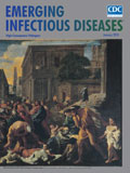
Of Rats and Men: Poussin’s Plague at Ashdod [PDF - 1.41 MB - 2 pages]
| EID | Asensi V, Fierer J. Of Rats and Men: Poussin’s Plague at Ashdod. Emerg Infect Dis. 2018;24(1):186-187. https://doi.org/10.3201/eid2401.ac2401 |
|---|---|
| AMA | Asensi V, Fierer J. Of Rats and Men: Poussin’s Plague at Ashdod. Emerging Infectious Diseases. 2018;24(1):186-187. doi:10.3201/eid2401.ac2401. |
| APA | Asensi, V., & Fierer, J. (2018). Of Rats and Men: Poussin’s Plague at Ashdod. Emerging Infectious Diseases, 24(1), 186-187. https://doi.org/10.3201/eid2401.ac2401. |


