Volume 10, Number 2—February 2004
THEME ISSUE
2004 SARS Edition
Laboratory Study
Ultrastructural Characterization of SARS Coronavirus
Figure 5
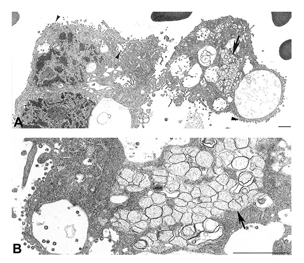
Figure 5. Ultrastructural characteristics of a bronchial alveolar lavage (BAL) from a patient with severe acute respiratory syndrome. A) Infected cells from a BAL specimen, showing numerous cytoplasmic and extracellular virions (arrowheads). Note the region of double-membrane vesicles (arrow), a common feature of coronavirus-infected cells. B) At higher magnification, double-membrane vesicles (arrow) are shown to contain diffuse, granular material. Bars, 1 μm.
Page created: January 31, 2011
Page updated: January 31, 2011
Page reviewed: January 31, 2011
The conclusions, findings, and opinions expressed by authors contributing to this journal do not necessarily reflect the official position of the U.S. Department of Health and Human Services, the Public Health Service, the Centers for Disease Control and Prevention, or the authors' affiliated institutions. Use of trade names is for identification only and does not imply endorsement by any of the groups named above.