Skip directly to site content Skip directly to page options Skip directly to A-Z link Skip directly to A-Z link Skip directly to A-Z link
Volume 10, Number 6—June 2004
Research

Candida parapsilosis Characterization in an Outbreak Setting

Duncan M. Kuhn*1, Pranab K. Mukherjee*, Thomas A. Clark†, Claude Pujol‡, Jyotsna Chandra*, Rana A. Hajjeh†, David W. Warnock†, David R. Soll‡, and Mahmoud A. Ghannoum*Comments to Author 
Author affiliations: *University Hospitals of Cleveland and Case Western Reserve University, Cleveland, Ohio, USA; †Centers for Disease Control and Prevention, Atlanta, Georgia, USA; ‡University of Iowa, Iowa City, Iowa, USA; 1Current affiliation: Division of Pulmonary and Critical Care, University of Washington Hospitals, Seattle, WA 98195, USA.

Main Article

Figure 5

Secretory aspartic protease (SAP) expression by Candida parapsilosis clinical isolates. Panel A shows representative sodium dodecyl sulfate–polyacrylamide gel electrophoresis of various C. parapsilosis isolates. M, molecular weight marker lane; BSA, bovine serum albumin alone; other lanes show number of isolate; and +, supernatant plus protease inhibitor cocktail. Protease activity is evident from the appearance of lower molecular weight bands representing cleavage products. Thick arrow indicate

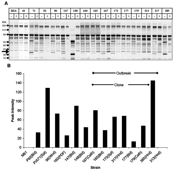
Figure 5. Secretory aspartic protease (SAP) expression by Candida parapsilosis clinical isolates. Panel A shows representative sodium dodecyl sulfate–polyacrylamide gel electrophoresis of various C. parapsilosis isolates. M, molecular weight marker lane; BSA, bovine serum albumin alone; other lanes show number of isolate; and +, supernatant plus protease inhibitor cocktail. Protease activity is evident from the appearance of lower molecular weight bands representing cleavage products. Thick arrow indicates the 20-kDa protein appearing after protease digestion. (For details of methods used see text.) Panel B shows densitometric scanning analysis of SAP activity. Strains 177 and 179 were included to demonstrate the heterogeneity in SAP production within the clonal strains. Cath, catheter; Bld, bloodstream; Spt, sputum; Hnd, hand; Wnd, wound; Pdf, peritoneal dialysis fluid.

Main Article

Page created: February 22, 2011
Page updated: February 22, 2011
Page reviewed: February 22, 2011
The conclusions, findings, and opinions expressed by authors contributing to this journal do not necessarily reflect the official position of the U.S. Department of Health and Human Services, the Public Health Service, the Centers for Disease Control and Prevention, or the authors' affiliated institutions. Use of trade names is for identification only and does not imply endorsement by any of the groups named above.
file_external