Skip directly to site content Skip directly to page options Skip directly to A-Z link Skip directly to A-Z link Skip directly to A-Z link
Volume 14, Number 1—January 2008
Dispatch

Protochlamydia naegleriophila as Etiologic Agent of Pneumonia

Nicola Casson*, Rolf Michel†, Karl-Dieter Müller, John David Aubert*, and Gilbert Greub*Comments to Author 
Author affiliations: *University Hospital Center and University of Lausanne, Lausanne, Switzerland; †Central Institute of the Federal Armed Forces Medical Services, Koblenz, Germany; ‡Institut für Medizinische Mikrobiologie der Universität Essen, Essen, Germany;

Main Article

Figure 1

Transmission electron microscopy of Protochlamydia naegleriophila. A) Naegleria lovaniensis trophozoite after transfer of endocytobionts; strain KNic (p) from the original host strain showing 15 coccoid bacteria distributed randomly within the cytoplasm of the host ameba. N, nucleus; en, endosome (karyosome) within the nucleus; v, empty food vacuoles. Magnification ×10,500; bar = 1 μm. B) N. lovaniensis trophozoite jammed with numerous endoparasitic stages of Pr. naegleriophila. Magnification ×16,800; bar = 1 μm. C) Enlarged detail of N. lovaniensis trophozoite with intracytoplasmic stages of Pr. naegleriophila. Some stages show binary fission indicated by the fission furrow (arrows). The endoparasites have a wrinkled gram-negative outer membrane rendering a spiny appearance to the endoparasites. Signs of damage are obvious within the cytoplasm of the host ameba. mi, mitochondria. Magnification ×43,500; bar = 0.5 μm. D) Pr. naegleriophila within vacuoles of Acanthamoebae castellanii ameba 2 days postinfection. Elementary bodies (eb) and reticulate bodies (rb) are visible. Elementary bodies harbor a smooth membrane compared with the reticulate bodies, which have a spiny shape. Magnification ×10,000; bar = 2 μm. E) Enlarged detail of A. castellanii trophozoite with intracytoplasmic stages of Pr. naegleriophila 3 days postinfection. Binary fission is observed (di). Magnification ×20,000; bar = 1 μm. F) Crescent body (arrow) within A. castellanii observed 3 days postinfection. Magnification ×20,000; bar = 1 μm.

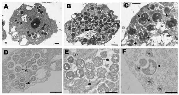
Figure 1. Transmission electron microscopy of Protochlamydia naegleriophila. A) Naegleria lovaniensis trophozoite after transfer of endocytobionts; strain KNic (p) from the original host strain showing 15 coccoid bacteria distributed randomly within the cytoplasm of the host ameba. N, nucleus; en, endosome (karyosome) within the nucleus; v, empty food vacuoles. Magnification ×10,500; bar = 1 μm. B) N. lovaniensis trophozoite jammed with numerous endoparasitic stages of Pr. naegleriophila. Magnification ×16,800; bar = 1 μm. C) Enlarged detail of N. lovaniensis trophozoite with intracytoplasmic stages of Pr. naegleriophila. Some stages show binary fission indicated by the fission furrow (arrows). The endoparasites have a wrinkled gram-negative outer membrane rendering a spiny appearance to the endoparasites. Signs of damage are obvious within the cytoplasm of the host ameba. mi, mitochondria. Magnification ×43,500; bar = 0.5 μm. D) Pr. naegleriophila within vacuoles of Acanthamoebae castellanii ameba 2 days postinfection. Elementary bodies (eb) and reticulate bodies (rb) are visible. Elementary bodies harbor a smooth membrane compared with the reticulate bodies, which have a spiny shape. Magnification ×10,000; bar = 2 μm. E) Enlarged detail of A. castellanii trophozoite with intracytoplasmic stages of Pr. naegleriophila 3 days postinfection. Binary fission is observed (di). Magnification ×20,000; bar = 1 μm. F) Crescent body (arrow) within A. castellanii observed 3 days postinfection. Magnification ×20,000; bar = 1 μm.

Main Article

Page created: July 07, 2010
Page updated: July 07, 2010
Page reviewed: July 07, 2010
The conclusions, findings, and opinions expressed by authors contributing to this journal do not necessarily reflect the official position of the U.S. Department of Health and Human Services, the Public Health Service, the Centers for Disease Control and Prevention, or the authors' affiliated institutions. Use of trade names is for identification only and does not imply endorsement by any of the groups named above.
file_external