Skip directly to site content Skip directly to page options Skip directly to A-Z link Skip directly to A-Z link Skip directly to A-Z link
Volume 14, Number 3—March 2008
Dispatch

Dolphin Morbillivirus Epizootic Resurgence, Mediterranean Sea

Juan-Antonio Raga*, Ashley Banyard†Comments to Author , Mariano Domingo‡, Mandy Corteyn†, Marie-Françoise Van Bressem§, Mercedes Fernández*, Francisco-Javier Aznar*, and Thomas Barrett†
Author affiliations: *University of Valencia, Valencia, Spain; †Institute for Animal Health, Pirbright, UK; ‡Autonomous University of Barcelona, Bellaterra, Barcelona, Spain; §Museo de Delfines, Pucusana, Peru;

Main Article

Figure 2

Evidence concerning the identity of the morbillivirus infecting Mediterranean striped dolphins in 2007. A) Immunohistologic staining of lung tissue with a MoAb anti CDV-NP, counterstained with hematoxylin. Epithelial bronchiolar cells and cells in the bronchiolar lumen are positively stained. B) Phylogenetic analysis of the N1/N2 region of the morbillivirus N genes. The number indicated at each node represents the bootstrap value after 10,000 replicates. The evolutionary distances were computed with the Kimura 2-parameter method and are in the units of the number of base substitutions per site. The tree is drawn to scale, with branch lengths in the same units as those of the evolutionary distances used to infer the phylogenetic tree. Details of each isolate used for this study and accession numbers for each of the sequences are as follows: DMV 2007 isolate (EU124652), PMV G12 isolate (AY949833), PPRV Nigeria/75/1 Vaccine (X74443), PDV DK/88 strain (X75717), RPV RBOK vaccine strain (Z30697), DMV isolate (AJ608288), CDV Onderstepoort vaccine strain (AF305419), and MV Edmonston vaccine strain (AF266288). DMV, dolphin morbillivirus; PMV, porpoise morbillivirus; PPRV, peste des petits ruminants virus; PDV, phocine distemper virus; RPV RBOK, rinderpest virus RBOK strain; CDV, canine distemper virus; MV, measles virus.

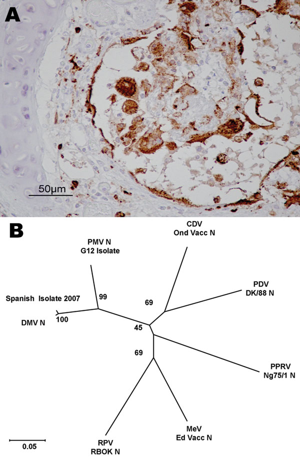
Figure 2. Evidence concerning the identity of the morbillivirus infecting Mediterranean striped dolphins in 2007. A) Immunohistologic staining of lung tissue with a MoAb anti CDV-NP, counterstained with hematoxylin. Epithelial bronchiolar cells and cells in the bronchiolar lumen are positively stained. B) Phylogenetic analysis of the N1/N2 region of the morbillivirus N genes. The number indicated at each node represents the bootstrap value after 10,000 replicates. The evolutionary distances were computed with the Kimura 2-parameter method and are in the units of the number of base substitutions per site. The tree is drawn to scale, with branch lengths in the same units as those of the evolutionary distances used to infer the phylogenetic tree. Details of each isolate used for this study and accession numbers for each of the sequences are as follows: DMV 2007 isolate (EU124652), PMV G12 isolate (AY949833), PPRV Nigeria/75/1 Vaccine (X74443), PDV DK/88 strain (X75717), RPV RBOK vaccine strain (Z30697), DMV isolate (AJ608288), CDV Onderstepoort vaccine strain (AF305419), and MV Edmonston vaccine strain (AF266288). DMV, dolphin morbillivirus; PMV, porpoise morbillivirus; PPRV, peste des petits ruminants virus; PDV, phocine distemper virus; RPV RBOK, rinderpest virus RBOK strain; CDV, canine distemper virus; MV, measles virus.

Main Article

Page created: July 07, 2010
Page updated: July 07, 2010
Page reviewed: July 07, 2010
The conclusions, findings, and opinions expressed by authors contributing to this journal do not necessarily reflect the official position of the U.S. Department of Health and Human Services, the Public Health Service, the Centers for Disease Control and Prevention, or the authors' affiliated institutions. Use of trade names is for identification only and does not imply endorsement by any of the groups named above.
file_external