Skip directly to site content Skip directly to page options Skip directly to A-Z link Skip directly to A-Z link Skip directly to A-Z link
Volume 14, Number 4—April 2008
Research

Retrospective Analysis of Monkeypox Infection

Melissa E. Dubois* and Mark K. Slifka*Comments to Author 
Author affiliations: *Oregon Health and Science University, Beaverton, Oregon, USA;

Main Article

Figure 1

Diagnosis of monkeypox by postadsorption ELISA. Plasma samples were obtained from monkeypox-immune persons (2–30 months postinfection), vaccinia-immune persons (2–4 months postinfection), or uninfected orthopoxvirus-naive persons and tested on ELISA plates coated with inactivated monkeypox antigen. A) A representative monkeypox-specific ELISA with plasma samples from an unvaccinated monkeypox-infected person (MPV), a previously vaccinated (i.e., vaccinia-immune) monkeypox-infected person (VV-MPV), a vaccinia-immune person (VV), a vaccinia-immune person who was revaccinated with vaccinia (VV-VV), and an uninfected orthopoxvirus-naive person (OPV-naive). Plasma was not preadsorbed (∅, gray bars), preadsorbed with inactivated vaccinia antigen (black bars), or preadsorbed with inactivated monkeypox antigen (white bars) before ELISA on monkeypox-coated plates. Numbers above bars refer to differences in postadsorption MPV ELISA titers after adsorption with vaccinia antigen compared with adsorption with monkeypox antigen. Plasma from 1 orthopoxvirus-naive person (representative of n = 12) was not preadsorbed with viral antigen because it was seronegative (<100 ELISA units) and below our detection limit (dashed horizontal line). B) Plasma samples from monkeypox-infected persons (•, n = 13), vaccinia-immune monkeypox-infected persons (■, n = 8), vaccinia-immune persons (▲, n = 10), and revaccinated vaccinia-immune persons (▼, n = 10) were tested by postadsorption ELISA. Data show fold-differences of monkeypox antibody titers after adsorption with vaccinia antigen compared with adsorption with monkeypox antigen. Dashed horizontal line indicates a diagnostic cutoff indicative of a positive result, which was determined as a postadsorption difference score of >2.5. *Denotes results of plasma samples obtained from persons with clinically inapparent monkeypox infection.

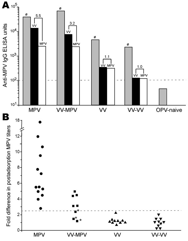
Figure 1. Diagnosis of monkeypox by postadsorption ELISA. Plasma samples were obtained from monkeypox-immune persons (2–30 months postinfection), vaccinia-immune persons (2–4 months postinfection), or uninfected orthopoxvirus-naive persons and tested on ELISA plates coated with inactivated monkeypox antigen. A) A representative monkeypox-specific ELISA with plasma samples from an unvaccinated monkeypox-infected person (MPV), a previously vaccinated (i.e., vaccinia-immune) monkeypox-infected person (VV-MPV), a vaccinia-immune person (VV), a vaccinia-immune person who was revaccinated with vaccinia (VV-VV), and an uninfected orthopoxvirus-naive person (OPV-naive). Plasma was not preadsorbed (∅, gray bars), preadsorbed with inactivated vaccinia antigen (black bars), or preadsorbed with inactivated monkeypox antigen (white bars) before ELISA on monkeypox-coated plates. Numbers above bars refer to differences in postadsorption MPV ELISA titers after adsorption with vaccinia antigen compared with adsorption with monkeypox antigen. Plasma from 1 orthopoxvirus-naive person (representative of n = 12) was not preadsorbed with viral antigen because it was seronegative (<100 ELISA units) and below our detection limit (dashed horizontal line). B) Plasma samples from monkeypox-infected persons (•, n = 13), vaccinia-immune monkeypox-infected persons (■, n = 8), vaccinia-immune persons (▲, n = 10), and revaccinated vaccinia-immune persons (▼, n = 10) were tested by postadsorption ELISA. Data show fold-differences of monkeypox antibody titers after adsorption with vaccinia antigen compared with adsorption with monkeypox antigen. Dashed horizontal line indicates a diagnostic cutoff indicative of a positive result, which was determined as a postadsorption difference score of >2.5. *Denotes results of plasma samples obtained from persons with clinically inapparent monkeypox infection.

Main Article

Page created: July 13, 2010
Page updated: July 13, 2010
Page reviewed: July 13, 2010
The conclusions, findings, and opinions expressed by authors contributing to this journal do not necessarily reflect the official position of the U.S. Department of Health and Human Services, the Public Health Service, the Centers for Disease Control and Prevention, or the authors' affiliated institutions. Use of trade names is for identification only and does not imply endorsement by any of the groups named above.
file_external