Volume 16, Number 4—April 2010
Research
Influenza A Strain-Dependent Pathogenesis in Fatal H1N1 and H5N1 Subtype Infections of Mice
Figure 4
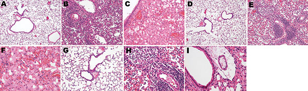
Figure 4. Photomicrographs of the lung sections of influenza A virus (H1N1)– and (H5N1)–infected mice at endpoint (hematoxylin and eosin stain). Dramatically different histopathologic signatures are observed, with either a mostly cellular reaction (H1N1) or a mostly humoral reaction (H5N1). Panels A, D, and G: 3 views of vehicle-infected lungs (original magnification ×100). Panels B and E, subtype H1N1: Dense granulocytic and lymphocytic cell infiltrates in the interstitium and around vessels and airways with focally denuded lamina propria due to epithelial necrosis and desquamation (original magnification ×100). Panel C, subtype H5N1: Airway epithelium is intact; note the striking difference in the number of infiltrated inflammatory cells between subtypes H1N1- and H5N1-infected lungs. Dramatic congestion of the vessels is visible, with extensive interstitial and alveolar edema (original magnification ×100). Panel F, subtype H5N1: Alveoli are completely filled with edema and hemorrhages; cellular infiltrates are conspicuously absent (original magnification ×200). Panel H, subtype H1N1: An airway with a totally denuded lamina propria is shown (top, left), with its lumen filled with granulocytic and lymphocytic exsudate (original magnification ×200). A prominent periarteriolar lymphocytic cuff is visible (bottom right). Panel I, subtype H5N1: Moderate inflammatory cell infiltrate, with no cuffing of any airway or vessel; an airway with a still intact epithelium is shown, located just beside a vessel with dramatic peripheral edema (original magnification ×200).