Volume 3, Number 1—March 1997
Synopsis
Surface Antigens of the Syphilis Spirochete and Their Potential as Virulence Determinants
Figure 2
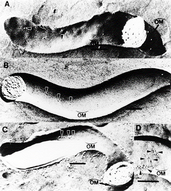
Figure 2.
Freeze-fracture electron microscopy of T. pallidum subsp. pallidum demonstrating TROMP aggregation. Concave
Page created: December 21, 2010
Page updated: December 21, 2010
Page reviewed: December 21, 2010
The conclusions, findings, and opinions expressed by authors contributing to this journal do not necessarily reflect the official position of the U.S. Department of Health and Human Services, the Public Health Service, the Centers for Disease Control and Prevention, or the authors' affiliated institutions. Use of trade names is for identification only and does not imply endorsement by any of the groups named above.