Volume 19, Number 9—September 2013
Letter
Chorea and Tick-Borne Encephalitis, Poland
Joanna Zajkowska1, Anna Moniuszko1 , Piotr Czupryna, Wiesław Drozdowski, Waldemar Krupa, Katarzyna Guziejko, Maciej Kondrusik, Sambor Grygorczuk, and Slawomir Pancewicz
Figure
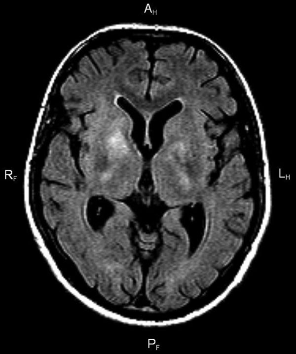
Figure. . . Fluid-attenuated inversion recovery magnetic resonance imaging of the brain of a 38-year-old man with tick-borne encephalitis and chorea. The image shows bilateral areas of hyperintensity in T2, affecting the nucleus caudate, internal capsule, and thalami.
1These authors contributed equally to this article.
Page created: August 15, 2013
Page updated: August 15, 2013
Page reviewed: August 15, 2013
The conclusions, findings, and opinions expressed by authors contributing to this journal do not necessarily reflect the official position of the U.S. Department of Health and Human Services, the Public Health Service, the Centers for Disease Control and Prevention, or the authors' affiliated institutions. Use of trade names is for identification only and does not imply endorsement by any of the groups named above.