Volume 20, Number 4—April 2014
Dispatch
Pathology of US Porcine Epidemic Diarrhea Virus Strain PC21A in Gnotobiotic Pigs
Figure 2
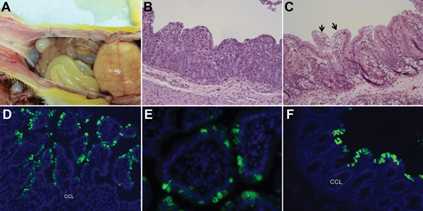
Figure 2. . Changes seen, by macroscopic examination, histologic examination, or immunofluorescence staining in the intestine of gnotobiotic pigs inoculated with porcine epidemic diarrhea virus (PEDV; US strain PC21A). A) Intestine of pig 1 at postinoculation hour (PIH) 30 (4–5 h after onset of clinical signs), showing thin and transparent intestinal walls (duodenum to colon) and extended stomach filled with curdled milk. B) Hematoxylin and eosin (H&E)–stained jejunum of pig 3 at PIH 46 (at onset of clinical signs), showing acute diffuse, severe atrophic jejunitis. Original magnification ×200. C) H&E-stained cecum of noninoculated pig 4 (which was exposed to inoculated pig 3 at PIH 0) at 120 h after onset of clinical signs. Acute diffuse, mild vacuolation of superficial epithelial cells (arrows) and subepithelial edema are seen. Original magnification ×200. D) Immunofluorescence staining of jejunum of pig 5 at PIH 67 (37–41 h after onset of clinical signs), indicating that the epithelial cells lining atrophied villi are positive for PEDV. Original magnification ×200. E) Immunofluorescence staining of jejunum of pig 3 at PIH 46 (at onset of clinical signs), showing localization of PEDV antigens in the cytoplasm of enterocytes. Original magnification ×600. F) Immunofluorescence staining of colon of pig 2 at PIH 72 (26–28 h after onset of clinical signs), showing large numbers of PEDV-positive cells. Original magnification ×200. CCL, crypt cell layer. Nuclei were stained with blue-fluorescent 4′, 6-diamidino-2-phenylindole, dihydrochloride.
1These authors were co-principal investigators.