Volume 13, Number 8—August 2007
Research
Venezuelan Equine Encephalitis Virus Infection of Cotton Rats
Anne-Sophie Carrara*, Lark L. Coffey*, Patricia V. Aguilar*, Abelardo C. Moncayo*, Amelia P.A. Travassos Da Rosa*, Marcio R.T. Nunes†, Robert B. Tesh*, and Nikos Vasilakis*
Figure 2
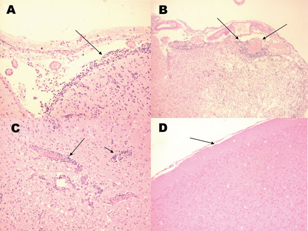
Figure 2. Histologic staining (hematoxylin and eosin) of Florida cotton rat tissues 9 days after intracranial inoculation with 3 log10 PFU of enzootic Venezuelan equine encephalitis virus (subtype IE). A) Inflammation of the meninges (arrows). B) Inflammation of the meninges and dilated blood vessels (arrows). C) Perivascular cuffing of blood vessels (arrow). D) Brain from a noninfected rat. (Magnification ×40.)
Page created: June 30, 2010
Page updated: June 30, 2010
Page reviewed: June 30, 2010
The conclusions, findings, and opinions expressed by authors contributing to this journal do not necessarily reflect the official position of the U.S. Department of Health and Human Services, the Public Health Service, the Centers for Disease Control and Prevention, or the authors' affiliated institutions. Use of trade names is for identification only and does not imply endorsement by any of the groups named above.