Volume 4, Number 2—June 1998
Synopsis
Enteroaggregative Escherichia coli
James P. Nataro* , Theodore Steiner†, and Richard L. Guerrant†
Figure 3
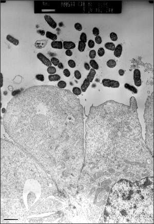
Figure 3. Effects of enteroaggregative E. coli strain 042 on T84 cells in culture. Polarized T84 monolayers were washed and inoculated with 106 CFU of 042 and allowed to coincubate at 37°C for 6 hrs. Transmission electron microscopy reveals adherence of bacteria to the apical surface of the T84 cells without internalization. The apical brush border is effaced; cells are ballooning and will eventually be extruded from the monolayer. (J.P. Nataro and S. Sears, unpub. data). Bar, 1 mm.
Page created: December 15, 2010
Page updated: December 15, 2010
Page reviewed: December 15, 2010
The conclusions, findings, and opinions expressed by authors contributing to this journal do not necessarily reflect the official position of the U.S. Department of Health and Human Services, the Public Health Service, the Centers for Disease Control and Prevention, or the authors' affiliated institutions. Use of trade names is for identification only and does not imply endorsement by any of the groups named above.