Volume 6, Number 3—June 2000
Research
Rhinosporidium seeberi: A Human Pathogen from a Novel Group of Aquatic Protistan Parasites
David N. Fredricks*† , Jennifer A. Jolley*, Paul W. Lepp*, Jon C. Kosek†, and David A. Relman*†
Figure 1
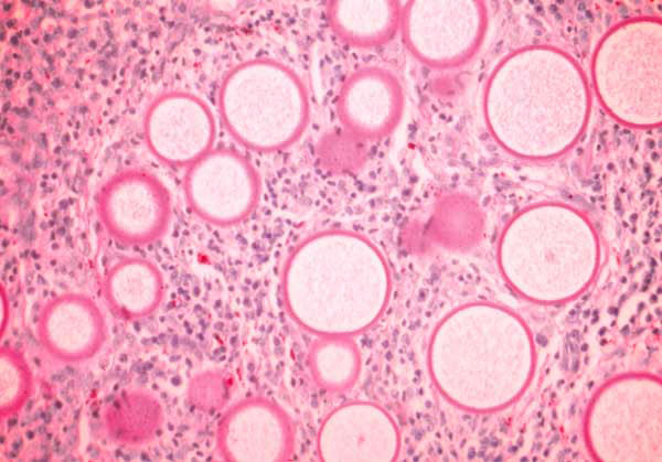
Figure 1. Histology of rhinosporidiosis. A formaldehyde-fixed section of human nasal polyp was stained with Periodic acid-Schiff (PAS) and visualized by bright-field microscopy at 400X magnification. The thick walls of immature R. seeberi trophocytes stain with PAS (pink), and the spherical organisms are surrounded by inflammatory cells.
Page created: December 16, 2010
Page updated: December 16, 2010
Page reviewed: December 16, 2010
The conclusions, findings, and opinions expressed by authors contributing to this journal do not necessarily reflect the official position of the U.S. Department of Health and Human Services, the Public Health Service, the Centers for Disease Control and Prevention, or the authors' affiliated institutions. Use of trade names is for identification only and does not imply endorsement by any of the groups named above.