Volume 12, Number 3—March 2006
Research
Pneumonic Plague Cluster, Uganda, 2004
Elizabeth M. Begier* , Gershim Asiki†, Zaccheus Anywaine†, Brook Yockey‡, Martin Schriefer‡, Philliam Aleti§, Asaph Ogen-Odoi§, J. Erin Staples*‡, Christopher Sexton‡, Scott Bearden‡, and Jacob L. Kool‡
Figure 2
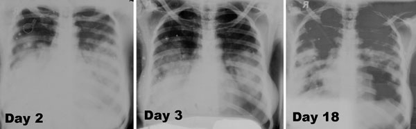
Figure 2. Three serial frontal chest radiographs from surviving caregiver B2 with primary pneumonic plague obtained on illness days 2, 3, and 18 showing bilateral lower lung zone predominant airspace disease associated with bilateral (right > left) pleural effusions. The radiographs have artifacts related to hand-dipping of the films, which account for multiple densities that move between images and the areas of apparent lucency.
Page created: January 27, 2012
Page updated: January 27, 2012
Page reviewed: January 27, 2012
The conclusions, findings, and opinions expressed by authors contributing to this journal do not necessarily reflect the official position of the U.S. Department of Health and Human Services, the Public Health Service, the Centers for Disease Control and Prevention, or the authors' affiliated institutions. Use of trade names is for identification only and does not imply endorsement by any of the groups named above.