Skip directly to site content Skip directly to page options Skip directly to A-Z link Skip directly to A-Z link Skip directly to A-Z link
Volume 14, Number 12—December 2008
Letter

Parachlamydia acanthamoebae Infection and Abortion in Small Ruminants

Silke Ruhl, Genevieve Goy, Nicola Casson, Rudolf Thoma, Andreas Pospischil, Gilbert Greub, and Nicole BorelComments to Author 
Author affiliations: University of Zurich, Zurich, Switzerland (S. Ruhl, A. Pospischil, N. Borel); University of Lausanne, Lausanne, Switzerland (G. Goy, N. Casson, G. Greub); Cantonal Laboratory of Veterinary Bacteriology, Chur, Switzerland (R. Thoma)

Main Article

Figure

A) Sheep placenta positive by real-time PCR and immunohistochemistry for Parachlamydia spp. and Chlamydiaceae. Chlamydophila abortus was identified by ArrayTube Microarray. Necrotizing placentitis and vasculitis are shown (hematoxylin and eosin stain; magnification ×200). B) Fetal lung of the sheep abortion specimen positive by real-time PCR and immunohistochemical tests for Parachlamydia spp. and Chlamydiaceae; interstitial pneumonia is shown (hematoxylin and eosin stain; magnification ×200). C) Fetal lung that was positive by real-time PCR and immunohistochemical testing for Parachlamydia spp. Positive granular material can be seen within the lung tissue. Antigen detection (immunohistochemistry) was carried out with a polyclonal antibody directed against Parachlamydia spp. 3-amino-9-ethylcarbazole/peroxidase method (hematoxylin counterstain; magnification ×200). D) Double immunohistochemical labeling of the sheep placenta that was positive by real-time PCR and immunohistochemical tests for Chlamydiaceae and Parachlamydia spp. The simultaneous presence of Chlamydiaceae and Parachlamydia spp. granular reaction is shown within necrotic trophoblastic epithelium and neutrophilic exudate (diaminobenzidine/AEC/peroxidase method, hematoxylin counterstain; magnification ×1,000).

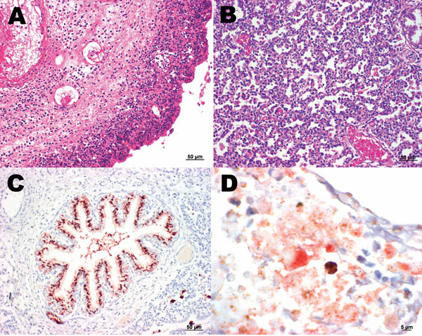
Figure. A) Sheep placenta positive by real-time PCR and immunohistochemistry for Parachlamydia spp. and Chlamydiaceae. Chlamydophila abortus was identified by ArrayTube Microarray. Necrotizing placentitis and vasculitis are shown (hematoxylin and eosin stain; magnification ×200). B) Fetal lung of the sheep abortion specimen positive by real-time PCR and immunohistochemical tests for Parachlamydia spp. and Chlamydiaceae; interstitial pneumonia is shown (hematoxylin and eosin stain; magnification ×200). C) Fetal lung that was positive by real-time PCR and immunohistochemical testing for Parachlamydia spp. Positive granular material can be seen within the lung tissue. Antigen detection (immunohistochemistry) was carried out with a polyclonal antibody directed against Parachlamydia spp. 3-amino-9-ethylcarbazole/peroxidase method (hematoxylin counterstain; magnification ×200). D) Double immunohistochemical labeling of the sheep placenta that was positive by real-time PCR and immunohistochemical tests for Chlamydiaceae and Parachlamydia spp. The simultaneous presence of Chlamydiaceae and Parachlamydia spp. granular reaction is shown within necrotic trophoblastic epithelium and neutrophilic exudate (diaminobenzidine/AEC/peroxidase method, hematoxylin counterstain; magnification ×1,000).

Main Article

Page created: July 22, 2010
Page updated: July 22, 2010
Page reviewed: July 22, 2010
The conclusions, findings, and opinions expressed by authors contributing to this journal do not necessarily reflect the official position of the U.S. Department of Health and Human Services, the Public Health Service, the Centers for Disease Control and Prevention, or the authors' affiliated institutions. Use of trade names is for identification only and does not imply endorsement by any of the groups named above.
file_external