Volume 17, Number 1—January 2011
Dispatch
Identification of Rickettsial Infections by Using Cutaneous Swab Specimens and PCR
Yassina Bechah, Cristina Socolovschi, and Didier Raoult
Figure 1
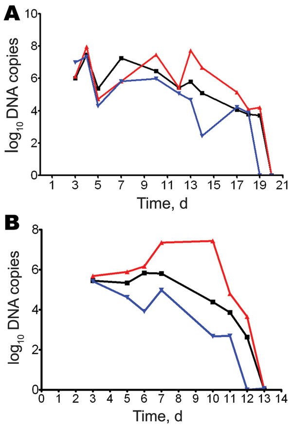
Figure 1. Molecular detection of Rickettsia spp. in swabs of skin lesions, Marseille, France. Guinea pigs were infected intradermally with different Rickettsia spp., and skin eschar swab specimens were obtained when lesions appeared. Samples (2 ± 1 mg) were tested, and DNA was extracted in a final volume of 100 μL. Number of rickettsial DNA copies was determined by quantitative PCR until day 20 postinfection for R. akari (black line), R. conorii (red line), and R. rhipicephali (blue line) (A) and until day 13 postinfection for R. africae (black line), Orientia tsutsugamushi (red line), and R. parkeri (blue line) (B). Values are copies of citrate synthase A gene/5 μL swab extract.
Page created: July 08, 2011
Page updated: July 08, 2011
Page reviewed: July 08, 2011
The conclusions, findings, and opinions expressed by authors contributing to this journal do not necessarily reflect the official position of the U.S. Department of Health and Human Services, the Public Health Service, the Centers for Disease Control and Prevention, or the authors' affiliated institutions. Use of trade names is for identification only and does not imply endorsement by any of the groups named above.