Volume 17, Number 11—November 2011
Dispatch
Molecular Subtyping in Cholera Outbreak, Laos, 2010
Abstract
A cholera outbreak in Laos in July 2010 involved 237 cases, including 4 deaths. Molecular subtyping indicated relatedness between the Vibrio cholerae isolates in this and in a 2007 outbreak, uncovering a clonal group of V. cholerae circulating in the Mekong basin. Our finding suggests the subtyping methods will affect this relatedness.
Cholera is a major public health concern in countries where access to safe water and adequate sanitation cannot be guaranteed for all residents. Vibrio cholerae serogroups O1 and O139 are the causative agents of cholera (1). A major virulence factor is cholera toxin (Ctx) encoded by the ctxAB gene and located on the Ctx prophage. V. cholerae O1 is classified into 2 biotypes, classical and El Tor. The El Tor biotype is responsible for the ongoing seventh pandemic of cholera (2). Since the early 1990s, the El Tor variant strains, which are biotypes of El Tor but carry the classical type of ctxB, have emerged and prevail in multiple regions where cholera is endemic (1,3–6).
In July 2010, a cholera outbreak began in Attapeu Province in southern Laos along the Cambodian border. Onset dates were July 5–September 16. The outbreak spread to 17 villages of the province and involved 237 persons, including 4 who died. To isolate the suspected V. cholera colonies, we screened specimens on thiosulfate citrate bile salt sucrose agar with or without enrichment in alkaline peptone water. Suspected colonies were examined by conventional biochemical tests and PCR amplification of ctx (7,8). Of the 42 fecal specimens tested, 9 were culture positive. The isolates were toxigenic V. cholerae O1 serotype Ogawa with features of the El Tor variant, according to the ctxB-typing method of Morita et al. (9).
We analyzed the 9 V. cholerae isolates from the Attapeu outbreak. We performed pulsed-field gel electrophoresis (PFGE) according to the PulseNet protocol (10) and multilocus variable number tandem repeat analysis (MLVA) using the 7 loci, as described (5,11).
Figure
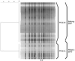
Figure. NotI-digested pulsed-field gel electrophoresis (PFGE) profiles of Vibrio cholerae isolates, Laos, 2010. The names of the profiles and the sources of the isolates are shown on the right. A dendrogram was...
The isolates of the Attapeu outbreak had almost indistinguishable PFGE profiles and MLVA repeat copy numbers. In PFGE analysis, 8 of the 9 isolates showed indistinguishable profiles (PFGE-A). The profile of the remaining isolate differed from the dominant isolates by 2 bands (PFGE-B) (Figure). In MLVA, 8 isolates showed the same MLVA type (MLVA-I), and 1 isolate showed another MLVA type that differed from the major MLVA type by being a single-locus variant of MLVA-I with only 1 locus and 1 repeat copy number (MLVA-II) (Table). Seven of the MLVA-I and 1 of the MLVA-II isolates showed the PFGE-A profile, and 1 of the MLVA-I isolates showed the PFGE-B profile. Although the source of contamination remains unknown, these results indicate that all isolates were indistinguishable from or similar to each other and that the outbreak could have been caused by a single source of contamination.
For comparison, we also examined 19 isolates from an outbreak that occurred in Xekong Province in 2007. These isolates also were toxigenic V. cholerae O1 serotype Ogawa of the El Tor variant (12). MLVA results clearly indicate that the isolates of the Attapeu outbreak in 2010 differed from those of the Xekong outbreak in 2007. The isolates from the Xekong outbreak comprised 3 MLVA types; 17 isolates were MLVA-III, 1 was MLVA-IV, and 1 was MLVA-V. MLVA-IV and MLVA-V were single-locus variants of MLVA-III (Table). Of the 7 loci tested, 3 or 4 displayed different repeat copy numbers than did those of the Attapeu and Xekong outbreaks. In PFGE analysis, however, the profiles were similar to each other; the isolates from the Xekong outbreak showed a PFGE-B profile (Figure).
These results suggest that strains with a specific PFGE type and the related strains have been circulating in the area for at least 3 years. Nguyen et al. suggested that another cholera outbreak in Vietnam that occurred from the end of 2007 to the beginning of 2008 was associated with the Xekong outbreak (13). Choi et al. also studied isolates from Vietnam in 2007 and 2008 by using MLVA, wherein they used 5 loci that are in common with those in this study (VC-1, -2, -6, -7, and -8) (14). The MLVA results obtained in our study indicated that the repeat copy numbers of the compatible loci of the Xekong outbreak isolates were the same as those of some of the isolates described in the study by Choi et al. This finding strongly suggests that the causative agents of the Xekong outbreak of Laos and the Vietnam outbreak in 2007–2008 were the same. Moreover, the strains were speculated to circulate widely in the Mekong basin, although the similarity between the PFGE profiles of the isolates from Laos and Vietnam remain to be studied.
Recently, another ctxB type of V. cholerae O1 biotype El Tor serotype Ogawa was reported in Orissa in eastern India (15). Representatives of the Xekong and Attapeu isolates also were subjected to sequence analysis of ctxB. The results showed that their ctxB sequences were identical with that of the original classical type, which suggests that the clonal group in the Mekong basin differs from the new Orissa type of V. cholerae in India.
Our study clearly indicates that the 2010 cholera outbreak at Attapeu was caused by 1 source of contamination. Furthermore, isolates from the Attapeu outbreak and the 2007 Xekong outbreak showed similar PFGE profiles, but they were differentiated by MLVA, consistent with their origin. This study suggests that PFGE analysis is useful for identifying the kinds of V. cholerae clones circulating in a specific geographic region and might be useful for determining a long-term framework of the region-specific V. cholerae because PFGE profiles are probably more stable than the MLVA types. By contrast, MLVA is useful for investigating and discriminating short-term individual outbreaks in a region. Another cholera outbreak in Cambodia in 2010 also might be related to the Attapeu outbreak. Combined use of both molecular subtyping methods would indicate the relatedness of cholera in the 2010 Cambodian outbreak and the others in the Mekong basin.
Dr Sithivong is head of the Bacteriology Unit of the National Center for Laboratory and Epidemiology, in Vientiane, Laos. Her research interests focus on the characteristics of bacterial agents causing diarrhea.
Acknowledgments
We thank Chanthavong Xayyasena and his team for technical assistance. We also thank all the donors who contributed financially to this study through the World Health Organization.
This study was partly supported by grants-in-aid from the Ministry of Health, Labour and Welfare of Japan (H20–Shinko–Ippan–013, H20–Shinko–Ippan–015, H21-Shokuhin-Ippan-005, H21-Shokuhin-Ippan-013, and H22-Shokuhin-Ippan-012) and from the Ministry of the Environment of Japan (Global Environment Research Fund, S-8).
References
- Raychoudhuri A, Mukhopadhyay AK, Ramamurthy T, Nandy RK, Takeda Y, Nair GB. Biotyping of Vibrio cholerae O1: time to redefine the scheme. Indian J Med Res. 2008;128:695–8.PubMedGoogle Scholar
- Nair GB, Qadri F, Holmgren J, Svennerholm AM, Safa A, Bhuiyan NA, Cholera due to altered El Tor strains of Vibrio cholerae O1 in Bangladesh. J Clin Microbiol. 2006;44:4211–3. DOIPubMedGoogle Scholar
- Morita M, Ohnishi M, Arakawa E, Yamamoto S, Nair GB, Matsushita S, Emergence and genetic diversity of El Tor Vibrio cholerae O1 that possess classical biotype ctxB among travel-associated cases of cholera in Japan. J Med Microbiol. 2010;59:708–12. DOIPubMedGoogle Scholar
- Safa A, Sultana J, Dac Cam P, Mwansa JC, Kong RY. Vibrio cholerae O1 hybrid El Tor strains, Asia and Africa. Emerg Infect Dis. 2008;14:987–8. DOIPubMedGoogle Scholar
- Abbott SL, Janda JM, Johnson JA. Farmer-III JJ. Vibrio and related organisms. In: Murray PR, Baron EJ, Jorgensen JH, Landry ML, Pfaller MA, editors. Manual of clinical microbiology. Vol. 1. 9th ed. Herndon (VA): ASM Press; 2007. p. 723–33.
- Nair GB, Shimada T, Kurazono H, Okuda J, Pal A, Karasawa T, Characterization of phenotypic, serological, and toxigenic traits of Vibrio cholerae O139 bengal. J Clin Microbiol. 1994;32:2775–9.PubMedGoogle Scholar
- Morita M, Ohnishi M, Arakawa E, Bhuiyan NA, Nusrin S, Alam M, Development and validation of a mismatch amplification mutation PCR assay to monitor the dissemination of an emerging variant of Vibrio cholerae O1 biotype El Tor. Microbiol Immunol. 2008;52:314–7. DOIPubMedGoogle Scholar
- Cooper KL, Luey CK, Bird M, Terajima J, Nair GB, Kam KM, Development and validation of a PulseNet standardized pulsed-field gel electrophoresis protocol for subtyping of Vibrio cholerae. Foodborne Pathog Dis. 2006;3:51–8. DOIPubMedGoogle Scholar
- Danin-Poleg Y, Cohen LA, Gancz H, Broza YY, Goldshmidt H, Malul E, Vibrio cholerae strain typing and phylogeny study based on simple sequence repeats. J Clin Microbiol. 2007;45:736–46. DOIPubMedGoogle Scholar
- Sithivong N, Izumiya H, Munnalath K, Phouthavane T, Chomlasak K, Sisavath L, Cholera outbreak, Laos, 2007. Emerg Infect Dis. 2010;16:745–6.PubMedGoogle Scholar
- Nguyen BM, Lee JH, Cuong NT, Choi SY, Hien NT, Anh DD, Cholera outbreaks caused by an altered Vibrio cholerae O1 El Tor biotype strain producing classical cholera toxin B in Vietnam in 2007 to 2008. J Clin Microbiol. 2009;47:1568–71. DOIPubMedGoogle Scholar
- Choi SY, Lee JH, Jeon YS, Lee HR, Kim EJ, Ansaruzzaman M, Multilocus variable-number tandem repeat analysis of Vibrio cholerae O1 El Tor strains harbouring classical toxin B. J Med Microbiol. 2010;59:763–9. DOIPubMedGoogle Scholar
- Kumar P, Jain M, Goel AK, Bhadauria S, Sharma SK, Kamboj DV, A large cholera outbreak due to a new cholera toxin variant of the Vibrio cholerae O1 El Tor biotype in Orissa, eastern India. J Med Microbiol. 2009;58:234–8. DOIPubMedGoogle Scholar
Figure
Table
Cite This Article1These authors contributed equally to this article.
Table of Contents – Volume 17, Number 11—November 2011
| EID Search Options |
|---|
|
|
|
|
|
|
Please use the form below to submit correspondence to the authors or contact them at the following address:
Hidemasa Izumiya, Department of Bacteriology I, National Institute of Infectious Diseases, Toyama 1-23-1,Shinjuku-ku, Tokyo 162-8640, Japan
Top