Volume 18, Number 4—April 2012
Letter
Tuberculosis Screening before Anti–Hepatitis C Virus Therapy in Prisons
Sergio Babudieri , Andrea Soddu, Monica Murino, Paola Molicotti, Alberto A. Muredda, Giordano Madeddu, Alessandro G. Fois, Stefania Zanetti, Pietro Pirina, and Maria Stella Mura
Figure
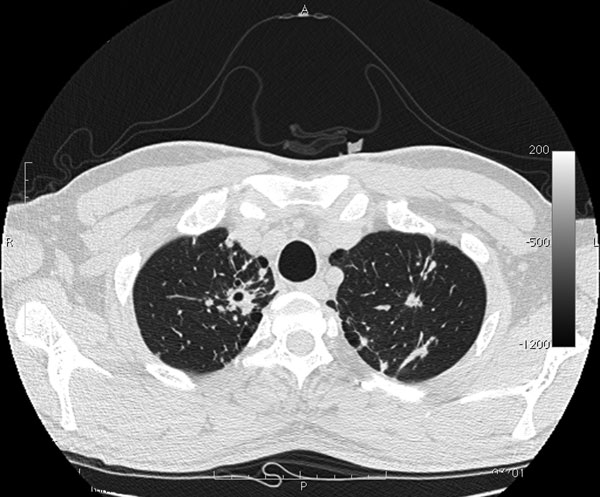
Figure. Computed tomography image of chest of patient with tuberculosis after anti–hepatitis C virus therapy. A parenchymal distortion 32 mm in diameter is shown in the upper right lung with initial central excavation 10 mm in diameter. Similar lesions 8 mm in diameter without central excavation are shown in the upper left lung.
Page created: March 16, 2012
Page updated: March 16, 2012
Page reviewed: March 16, 2012
The conclusions, findings, and opinions expressed by authors contributing to this journal do not necessarily reflect the official position of the U.S. Department of Health and Human Services, the Public Health Service, the Centers for Disease Control and Prevention, or the authors' affiliated institutions. Use of trade names is for identification only and does not imply endorsement by any of the groups named above.