Volume 21, Number 3—March 2015
Dispatch
Comparison of Porcine Epidemic Diarrhea Viruses from Germany and the United States, 2014
Dennis Hanke1, Maria Jenckel1, Anja Petrov, Mathias Ritzmann, Julia Stadler, Valerij Akimkin, Sandra Blome , Anne Pohlmann, Horst Schirrmeier, Martin Beer, and Dirk Höper
Figure 1
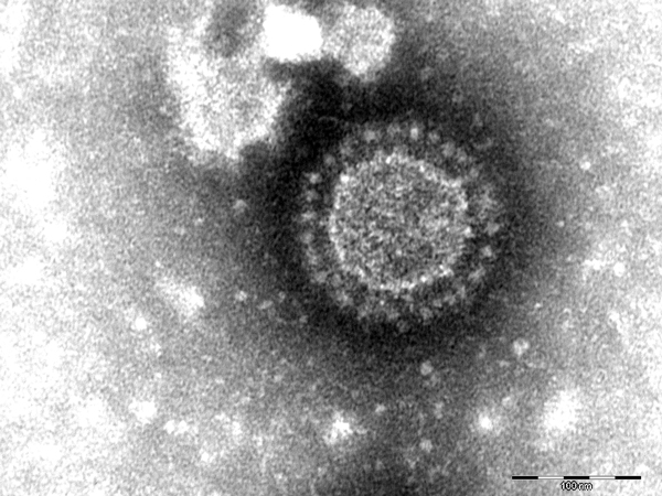
Figure 1. Porcine epidemic diarrhea virus particles seen by negative-stain electron microscopy of fecal samples. Negative staining with 1% phosphotungstic acid. Scale bar indicates 100 nm.
1These authors contributed equally to this article.
Page created: February 18, 2015
Page updated: February 18, 2015
Page reviewed: February 18, 2015
The conclusions, findings, and opinions expressed by authors contributing to this journal do not necessarily reflect the official position of the U.S. Department of Health and Human Services, the Public Health Service, the Centers for Disease Control and Prevention, or the authors' affiliated institutions. Use of trade names is for identification only and does not imply endorsement by any of the groups named above.