Skip directly to site content Skip directly to page options Skip directly to A-Z link Skip directly to A-Z link Skip directly to A-Z link
Volume 21, Number 5—May 2015
Letter

Fatal Balamuthia mandrillaris Meningoencephalitis in the Netherlands after Travel to The Gambia

Nadine A.M.E. van der Beek1, Carla van Tienen1, Jubi E. de Haan, Jeroen Roelfsema, Pieter J. Wismans, Perry J.J. van Genderen, Herve L. Tanghe, Rob M. Verdijk, Maarten J. Titulaer2, and Jaap J. van Hellemond2Comments to Author 
Author affiliations: Erasmus University Medical Centre, Rotterdam, the Netherlands (N.A.M.E. van der Beek, C. van Tienen, J.E. de Haan, H.L. Tanghe, R.M. Verdijk, M.J. Titulaer, J.J. van Hellemond); National Institute for Public Health and the Environment, Bilthoven, the Netherlands (J. Roelfsema); Harbor Hospital, Rotterdam (P.J. Wismans, P.J.J. van Genderen, J.J. van Hellemond)

Main Article

Figure

Postmortem pathologic findings for woman in the Netherlands who died of Balamuthia mandrillaris meningoencephalitis after returning from travel to The Gambia. A) Macroscopic coronal central section scan showing hemorrhagic necrotizing lesions of the subependymal, meningeal, and parenchymal areas of the parietotemporal lobes (circles and arrows). B) Low-power microscopic scan showing hemorrhagic necrotizing angiitis of the meningeal vessels (arrow) (original magnification ×25). C) Medium-power mi

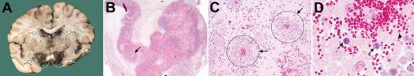
Figure. Postmortem pathologic findings for woman in the Netherlands who died of Balamuthia mandrillaris meningoencephalitis after returning from travel to The Gambia. A) Macroscopic coronal central section scan showing hemorrhagic necrotizing lesions of the subependymal, meningeal, and parenchymal areas of the parietotemporal lobes (circles and arrows). B) Low-power microscopic scan showing hemorrhagic necrotizing angiitis of the meningeal vessels (arrow) (original magnification ×25). C) Medium-power microscopic scan (original magnification ×200) showing perivascular trophozoite cuffing (arrows) and granulomatous inflammation. D) High-power microscopic scan (original magnification ×630) showing encysted amebae (arrows) and free trophozoites (arrowhead). Hematoxylin and eosin stains.

Main Article

1These first authors contributed equally to this article.

2These senior authors contributed equally to this article.

Page created: April 17, 2015
Page updated: April 17, 2015
Page reviewed: April 17, 2015
The conclusions, findings, and opinions expressed by authors contributing to this journal do not necessarily reflect the official position of the U.S. Department of Health and Human Services, the Public Health Service, the Centers for Disease Control and Prevention, or the authors' affiliated institutions. Use of trade names is for identification only and does not imply endorsement by any of the groups named above.
file_external