Skip directly to site content Skip directly to page options Skip directly to A-Z link Skip directly to A-Z link Skip directly to A-Z link
Volume 21, Number 9—September 2015
Letter

Fatal Accelerated Cirrhosis after Imported HEV Genotype 4 Infection

Ryan B. PerumpailComments to Author , Aijaz Ahmed, John P. Higgins, Samuel K. So, J. Lynn Cochran, Jan Drobeniuc, Tonya R. Mixson-Hayden, and Chong-Gee Teo
Author affiliations: Stanford University School of Medicine, Palo Alto, California, USA (R.B. Perumpail, A. Ahmed, J.P. Higgins, S.K. So); Birmingham Gastroenterology Associates, Birmingham, Alabama, USA (J.L. Cochran); Trinity Medical Center, Birmingham (J.L. Cochran); Centers for Disease Control and Prevention, Atlanta, Georgia, USA (J. Drobeniuc, T.R. Mixson-Hayden, C.-G. Teo)

Main Article

Figure

Serial histologic changes in liver of the patient who received a diagnosis of hepatitis E after a visit to Hong Kong in 2013 (A and B: at first biopsy; C and D: second biopsy; E and F: third biopsy. A) Mild mixed portal infiltration; minimal lobular inflammation; acidophil body present at upper right; and bile duct showing injury with lymphocytic infiltration (original magnification ×400). b) Mild portal inflammation; some interface activity; and portal tracts not showing increased fibrosity (or

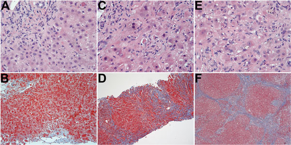
Figure. Serial histologic changes in liver of the patient who received a diagnosis of hepatitis E after a visit to Hong Kong in 2013 (A and B: at first biopsy; C and D: second biopsy; E and F: third biopsy. A) Mild mixed portal infiltration; minimal lobular inflammation; acidophil body present at upper right; and bile duct showing injury with lymphocytic infiltration (original magnification ×400). B) Mild portal inflammation; some interface activity; and portal tracts not showing increased fibrosity (original magnification ×200). C) Mononuclear infiltration of portal tract at upper right with bile duct/ductular infiltration and injury; lobular changes more severe, showing more inflammation, acidophil bodies and reactive nuclear change in hepatocytes with ballooning of some hepatocytes (original magnification ×400). D) Portal and lobular inflammation; and marked increase in fibrosis with bridging and regenerative nodule formation (original magnification ×100). E) Extensive lobular inflammation and reactive hepatocytic changes with nuclear enlargement, prominent nucleoli, and ballooning (original magnification ×400). F) Well-developed cirrhosis (original magnification ×40). Hematoxylin and eosin staining (A, C, E); Masson trichrome staining. (B, D, F).

Main Article

Page created: August 14, 2015
Page updated: August 14, 2015
Page reviewed: August 14, 2015
The conclusions, findings, and opinions expressed by authors contributing to this journal do not necessarily reflect the official position of the U.S. Department of Health and Human Services, the Public Health Service, the Centers for Disease Control and Prevention, or the authors' affiliated institutions. Use of trade names is for identification only and does not imply endorsement by any of the groups named above.
file_external