Volume 22, Number 12—December 2016
Dispatch
Horizontal Transmission of Chronic Wasting Disease in Reindeer
Figure 1
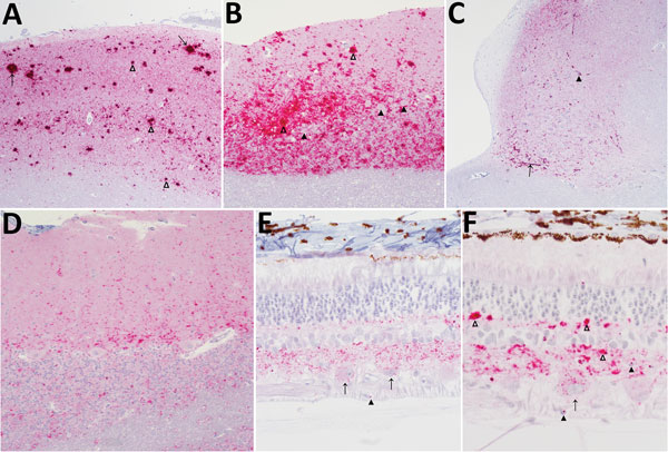
Figure 1. Immunohistochemical analysis for the prion protein showing scrapie prion protein (PrPSc) deposits in brains (A–D) and retinas (E, F) from reindeer (Rangifer tarandus tarandus) with chronic wasting disease. PrPSc immunodetection using the monoclonal antibody F99/97.6.1. A) Neocortex, showing prominent aggregated (open arrowheads) and plaque-like (arrows) deposits in reindeer no. 4. Original magnification ×5. B) Cerebellum, showing particulate immunoreactivity and aggregated deposits in reindeer no. 4. Note absence of intraneuronal immunoreactivity in Purkinje cells (solid arrowheads). Original magnification ×10 (open arrowheads). C) Brainstem at the level of the obex, showing prominent linear (arrow) and perineuronal (solid arrowhead) immunoreactivity in the dorsal motor nucleus of the vagus nerve in reindeer no. 21. Original magnification ×5. D) Cerebellum, punctate immunoreactivity in the molecular and granular layers and white matter in reindeer no. 12. Original magnification ×5. E) Intraneuronal immunoreactivity in retinal ganglion cells (arrows), punctate deposits in the inner and outer plexiform layers, scattered intramicroglial deposits (solid arrowheads) in reindeer no. 12. Original magnification ×40. F) Particulate to coalescing deposits in the inner and outer plexiform layers (open arrowheads), intraneuronal immunoreactivity in retinal ganglion cells (arrows), and scattered intramicroglial deposits (solid arrowheads) in reindeer no. 13. Original magnification ×40.
1Deceased.