Volume 22, Number 2—February 2016
Letter
Acute Colitis Caused by Helicobacter trogontum in Immunocompetent Patient
Fabien Dutasta1, Elia Samaha1, Nathalie Carayol, Jean-Marc Masse, Camille Bourillon, Clémence Richaud, Arthur Neuschwander, Hidayeth Rostane, Marie Lyse Parolini, Patrick Bruneval, Christophe Cellier, and Isabelle Podglajen
Figure
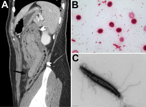
Figure. Computed tomographic image of patient with Helicobacter trogontum infection and micrographs of H. trogontum. A) Paramedian sagittal section of an abdominopelvic scan after injection of contrast medium in the portal phase, showing thickening of the transverse and right colon (white arrowheads) with tubular appearance and discrete thickening of the fat stranding (black arrow). B) Gram-stained blood culture smear. Original magnification ×1,000. C) Transmission electron micrograph of negatively stained H. trogontum showing bipolar flagella. Scale bar indicates 0.5 μm.
1These authors contributed equally to this article.
Page created: January 18, 2016
Page updated: January 18, 2016
Page reviewed: January 18, 2016
The conclusions, findings, and opinions expressed by authors contributing to this journal do not necessarily reflect the official position of the U.S. Department of Health and Human Services, the Public Health Service, the Centers for Disease Control and Prevention, or the authors' affiliated institutions. Use of trade names is for identification only and does not imply endorsement by any of the groups named above.